

Anti-BDNF Antibody (56572)


Data
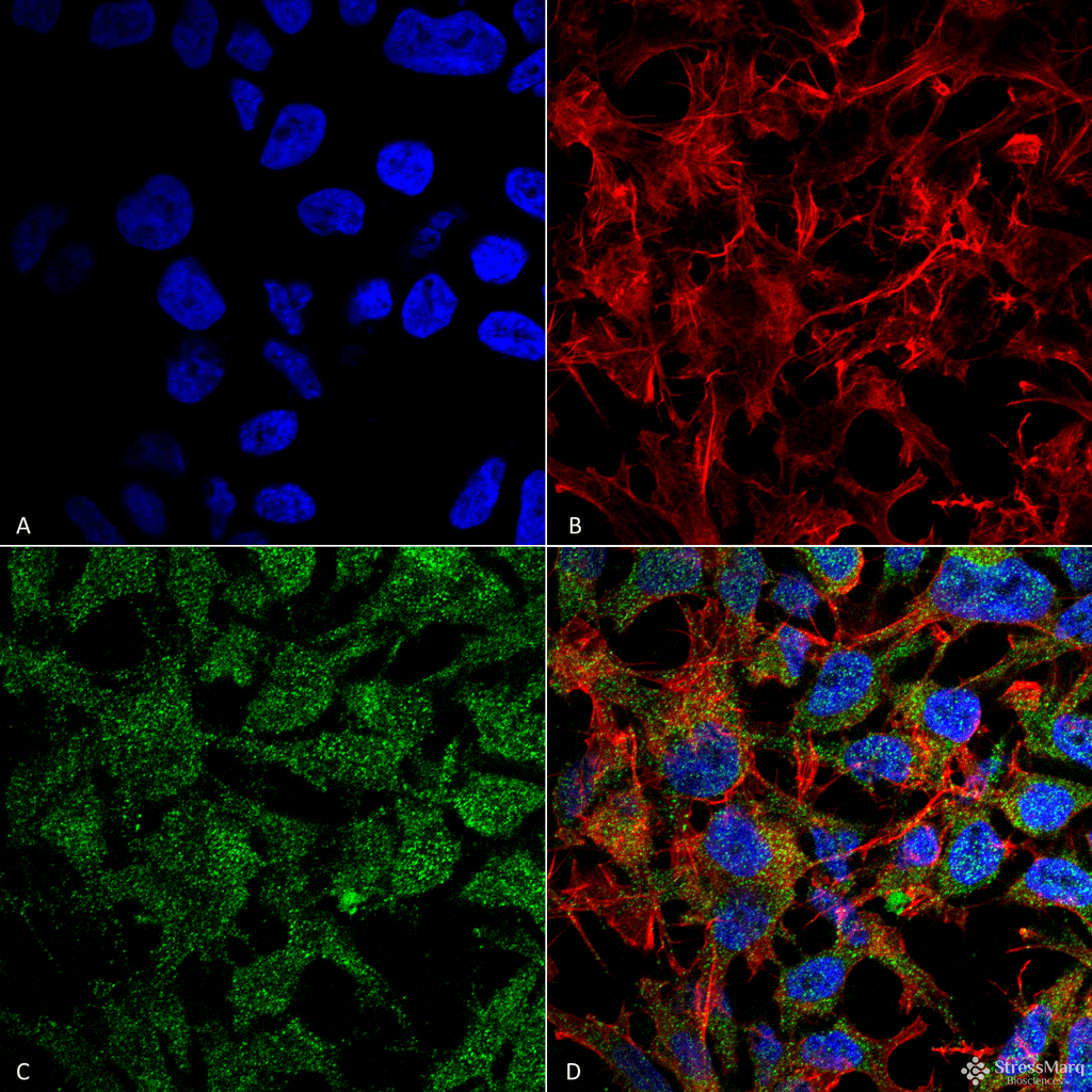 Immunocytochemistry/Immunofluorescence analysis using Rabbit Anti-BDNF Polyclonal Antibody (56572). Tissue: Neuroblastoma cell line (SK-N-BE). Species: Human. Fixation: 4% Formaldehyde for 15 min at RT. Primary Antibody: Rabbit Anti-BDNF Polyclonal Antibody (56572) at 1:100 for 60 min at RT. Secondary Antibody: Goat Anti-Rabbit ATTO 488 at 1:100 for 60 min at RT. Counterstain: Phalloidin Texas Red F-Actin stain; DAPI (blue) nuclear stain at 1:1000, 1:5000 for 60min RT, 5min RT. Localization: Secreted, Cytoplasm, Membrane-bound vesicle. Magnification: 60X. (A) DAPI (blue) nuclear stain (B) Phalloidin Texas Red F-Actin stain (C) BDNF Antibody (D) Composite.
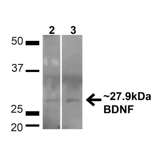 Western blot analysis of Human HeLa and HEK293T cell lysates showing detection of ~27.9 Kda BDNF protein using Rabbit Anti-BDNF Polyclonal Antibody (56572). Lane 1: MW Ladder. Lane 2: Human HeLa (20 µg). Lane 3: Human 293T (20 µg). Load: 20 µg. Block: 5% milk + TBST for 1 hour at RT. Primary Antibody: Rabbit Anti-BDNF Polyclonal Antibody (56572) at 1:1000 for 1 hour at RT. Secondary Antibody: Goat Anti-Rabbit: HRP at 1:2000 for 1 hour at RT. Color Development: TMB solution for 12 min at RT. Predicted/Observed Size: ~27.9 Kda.
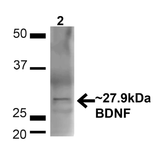 Western blot analysis of Mouse Brain showing detection of ~27.9 Kda BDNF protein using Rabbit Anti-BDNF Polyclonal Antibody (56572). Lane 1: MW Ladder. Lane 2: Mouse Brain (20 µg). Load: 20 µg. Block: 5% milk + TBST for 1 hour at RT. Primary Antibody: Rabbit Anti-BDNF Polyclonal Antibody (56572) at 1:1000 for 1 hour at RT. Secondary Antibody: Goat Anti-Rabbit: HRP at 1:2000 for 1 hour at RT. Color Development: TMB solution for 12 min at RT. Predicted/Observed Size: ~27.9 Kda.
 - -
- -
Antibody DetailsProduct DetailsReactive Species Human ⋅ Mouse Host Species Rabbit Immunogen Synthetic peptide corresponding to the N-terminus of human BDNF. Product Concentration 1.0 mg/ml Formulation PBS, pH 7.4, 50% glycerol, 0.09% sodium azide. State of Matter Liquid Product Preparation Purified by peptide immuno-affinity chromatography Storage and Handling This antibody is stable for at least one (1) year at -20°C. Regulatory Status For in vitro investigational use only. Not intended for therapeutic or diagnostic applications. Country of Origin USA Shipping Next Day 2-8°C Applications and Recommended Usage? Quality Tested by Leinco Immunoblotting: use at dilution of 1:1,000. A band of ~27-28kDa is detected.
Immunofluorescence: use at dilution of 1:100 These are recommended working dilutions. Endusers should determine optimal dilutions for their applications. Each investigator should determine their own optimal working dilution for specific applications. See directions on lot specific datasheets, as information may periodically change. DescriptionDescriptionSpecificity This antibody recognizes human and mouse BDNF. Background BDNF acts on neurons of the central nervous system and the peripheral nervous system supporting the survival of existing neurons and stimulating growth and differentiation of new neurons and synapses. In the brain, it is active in the hippocampus, cortex, and basal forebrain, areas vital to learning, memory, and higher thinking. It is also expressed in the retina, motor neurons, kidneys, saliva, and prostate. Function Important signaling molecule that activates signaling cascades downstream of NTRK2 (PubMed:11152678). During development, promotes the survival and differentiation of selected neuronal populations of the peripheral and central nervous systems. Participates in axonal growth, pathfinding and in the modulation of dendritic growth and morphology. Major regulator of synaptic transmission and plasticity at adult synapses in many regions of the CNS. The versatility of BDNF is emphasized by its contribution to a range of adaptive neuronal responses including long-term potentiation (LTP), long-term depression (LTD), certain forms of short-term synaptic plasticity, as well as homeostatic regulation of intrinsic neuronal excitability. {PubMed:11152678, PubMed:12553913, PubMed:29909994}.; [BDNF precursor form]: Important signaling molecule that activates signaling cascades downstream of NTRK2. Activates signaling cascades via the heterodimeric receptor formed by NGFR and SORCS2 (PubMed:24908487, PubMed:29909994). Signaling via NGFR and SORCS2 plays a role in synaptic plasticity and long-term depression (LTD). Binding to NGFR and SORCS2 promotes neuronal apoptosis. Promotes neuronal growth cone collapse (By similarity). {UniProtKB:P21237, PubMed:24908487, PubMed:29909994}. NCBI Gene Bank ID UniProt.org Research Area Neuroscience References & CitationsTechnical ProtocolsICC IF  Certificate of Analysis |
 Products are for research use only. Not for use in diagnostic or therapeutic procedures.