

Anti-CaMKII, phosphorylated, Antibody (56454)


Anti-CaMKII, phosphorylated, Antibody (56454)
Product No.: 56454
- -
- -
Clone 22B1 Target CaMKII, phosphorylated, Formats AvailableView All Product Type Monoclonal Alternate Names CaM kinase II subunitα, CaMK-II subunitα, EC 2.7.11.17 Isotype Mouse IgG1 Applications ELISA , IHC , IP , WB , ICC/IF |
Data
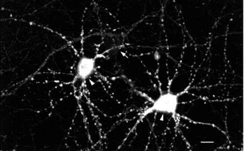 Immunocytochemistry/Immunofluorescence analysis using Mouse Anti-CaMKII Monoclonal Antibody, Clone 22B1 (56454). Tissue: dissociated hippocampal neurons. Species: Rat. Fixation: Cold 4% paraformaldehyde/0.2% glutaraldehyde in 0.1M sodium phosphate buffer. Primary Antibody: Mouse Anti-CaMKII Monoclonal Antibody (56454) at 1:1000 for 12 hours at 4°C. Secondary Antibody: FITC Goat Anti-Mouse IgG (green) at 1:50 for 30 minutes at RT. Magnification: 10X. Courtesy of: Mary Kennedy, Caltech.
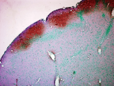 Immunohistochemistry analysis using Mouse Anti-CaMKII Monoclonal Antibody, Clone 22B1 (56454). Tissue: colon carcinoma. Species: Human. Fixation: Formalin. Primary Antibody: Mouse Anti-CaMKII Monoclonal Antibody (56454) at 1:5000 for 12 hours at 4°C. Secondary Antibody: Biotin Goat Anti-Mouse at 1:2000 for 1 hour at RT. Counterstain: Mayer Hematoxylin (purple/blue) nuclear stain at 200 µl for 2 minutes at RT. Magnification: 40x.
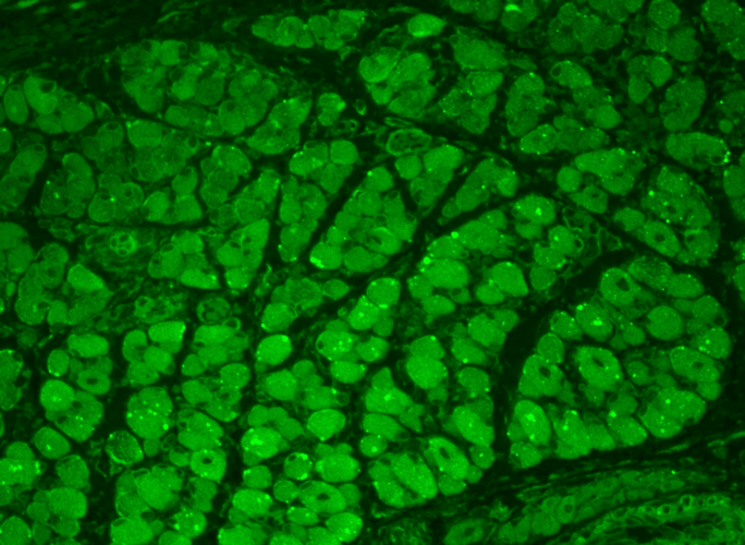 Immunohistochemistry analysis using Mouse Anti-CaMKII Monoclonal Antibody, Clone 22B1 (56454). Tissue: backskin. Species: Mouse. Fixation: Bouin’s Fixative and paraffin-embedded. Primary Antibody: Mouse Anti-CaMKII Monoclonal Antibody (56454) at 1:100 for 1 hour at RT. Secondary Antibody: FITC Goat Anti-Mouse (green) at 1:50 for 1 hour at RT. Localization: Muscle, hair follicle, epidermis. Backskin obtained from transgenic mice.
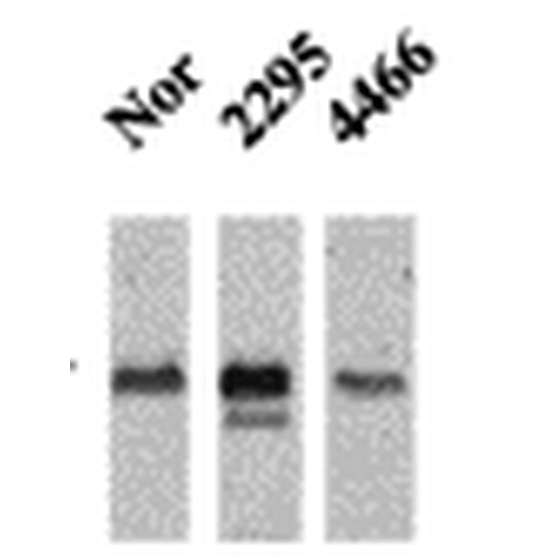 Western Blot analysis of Mouse Ventricle lysates showing detection of CaMKII protein using Mouse Anti-CaMKII Monoclonal Antibody, Clone 22B1 (56454). Primary Antibody: Mouse Anti-CaMKII Monoclonal Antibody (56454) at 1:1000. Analysis of CaMKII and NFAT phosphorylation in ventricles of 14 day old mice over-expressing CaMK.
 - -
- -
Antibody DetailsProduct DetailsReactive Species Mouse ⋅ Rat Host Species Mouse Immunogen Synthetic peptide based on sequence from rat/mouse CaMKII alpha (aa 281-294), thiophosphorylated at residue Thr286 conjugated to a protein carrier. Product Concentration Lot Specific Formulation PBS, pH 7.4. State of Matter Liquid Product Preparation Purified by Protein G affinity chromatography Storage and Handling This antibody is stable for at least one (1) year at -20°C. Avoid repeated freezing and
thawing. Regulatory Status For in vitro investigational use only. Not for
use in therapeutic or diagnostic procedures. Country of Origin USA Shipping Next Day 2-8°C Applications and Recommended Usage? Quality Tested by Leinco Immunoblotting: use at 1ug/mL. A band of ~50 kDa is detected.
Immunofluorescence: use at 10ug/mL. These are recommended concentrations. User should determine optimal concentrations for their application. Positive control: Rat brain tissue extract. Each investigator should determine their own optimal working dilution for specific applications. See directions on lot specific datasheets, as information may periodically change. DescriptionDescriptionSpecificity This antibody recognizes the
phosphorylated form of mouse and rat
CaMKII. It does not react with nonphosphorylated CaMKII. Background CaMKII, a member of the calcium / calmodulin-activated protein kinas family, functions in neural synapatic stimulation and T-cell receptor signaling. CaMKII is expressed in many tissues, but it is specifically found in the neurons of the forebrain, and its mRNA is found within the dendrites and the soma of the neuron. CaMKII in neurons consists of two subunits of 52 (alpha genes) and 60 kDa (beta genes). CaMKII has catalytic and regulatory domains as well as an ATP-binding domain and a consensus phosphorylation site. Binding of calcium / calmodulin to its regulatory domain releases its auto- inhibitory effect and activates the kinase. This kinase activation results in autophosphorylation at threonine 286. Autophosphorylation confers enhanced affinity of CaMKII for NMDA receptors in postsynaptic densities. Function Calcium/calmodulin-dependent protein kinase that functions autonomously after Ca(2+)/calmodulin-binding and autophosphorylation, and is involved in synaptic plasticity, neurotransmitter release and long-term potentiation. Member of the NMDAR signaling complex in excitatory synapses, it regulates NMDAR-dependent potentiation of the AMPAR and therefore excitatory synaptic transmission (PubMed:15312654). Regulates dendritic spine development. Also regulates the migration of developing neurons. Phosphorylates the transcription factor FOXO3 to activate its transcriptional activity (By similarity). Acts as a negative regulator of 2-arachidonoylglycerol (2-AG)-mediated synaptic signaling via modulation of DAGLA activity (By similarity). {UniProtKB:P11798, UniProtKB:Q9UQM7, PubMed:15312654}. NCBI Gene Bank ID UniProt.org Research Area Neuroscience References & CitationsTechnical Protocols    ICC/IF Certificate of Analysis |
Formats Available
 Products are for research use only. Not for use in diagnostic or therapeutic procedures.