

Anti-CD74 Antibody (56451)

Anti-CD74 Antibody (56451)
Product No.: 56451
- -
- -
Clone PIN1 Target CD74 Formats AvailableView All Product Type Monoclonal Alternate Names HLA-DR antigens-associated invariant chain, Ia antigen-associated invariant chain, Ii, CD antigen CD74 [Cleaved into: Class-II-associated invariant chain peptide Isotype Mouse IgG1 Applications FACS , IHC , IP , WB , FCM , ICC/IF |
Data
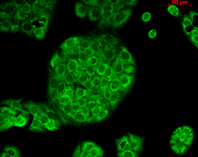 Immunocytochemistry/Immunofluorescence analysis using Mouse Anti-CD74 Monoclonal Antibody, Clone PIN 1.1 (56451). Tissue: HaCaT cells. Species: Human. Fixation: Cold 100% methanol for 10 minutes at -20°C. Primary Antibody: Mouse Anti-CD74 Monoclonal Antibody (56451) at 1:100 for 1 hour at RT. Secondary Antibody: FITC Goat Anti-Mouse (green) at 1:50 for 1 hour at RT. Localization: Cytoplasmic Staining.
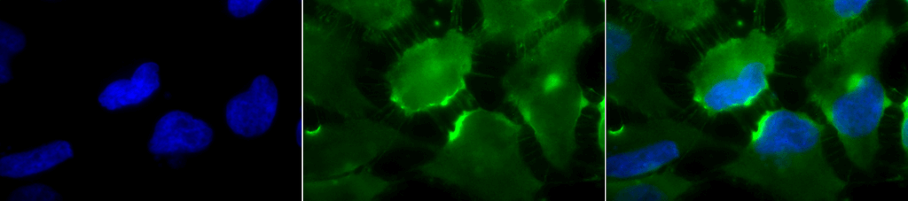 Immunocytochemistry/Immunofluorescence analysis using Mouse Anti-CD74 Monoclonal Antibody, Clone PIN 1.1 (56451). Tissue: Cervical cancer cell line (HeLa). Species: Human. Fixation: 2% Formaldehyde for 20 min at RT. Primary Antibody: Mouse Anti-CD74 Monoclonal Antibody (56451) at 1:100 for 12 hours at 4°C. Secondary Antibody: FITC Goat Anti-Mouse (green) at 1:200 for 2 hours at RT. Counterstain: DAPI (blue) nuclear stain at 1:40000 for 2 hours at RT. Localization: Cell membrane. Endoplasmic reticulum membrane. Golgi apparatus. Endosome. Lysosome. Magnification: 100x. (A) DAPI (blue) nuclear stain. (B) Anti-CD74 Antibody. (C) Composite.
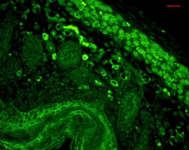 Immunohistochemistry analysis using Mouse Anti-CD74 Monoclonal Antibody, Clone PIN 1.1 (56451). Tissue: backskin. Species: Mouse. Fixation: Bouin’s Fixative and paraffin-embedded. Primary Antibody: Mouse Anti-CD74 Monoclonal Antibody (56451) at 1:100 for 1 hour at RT. Secondary Antibody: FITC Goat Anti-Mouse (green) at 1:50 for 1 hour at RT. Localization: Beautiful basal to suprabasal staining in epidermis, dermis, hair follicles and muscle.
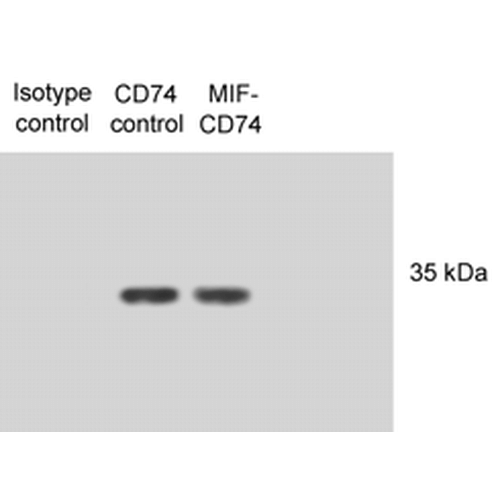 Western Blot analysis of Human N87 cell lysates showing detection of CD74 protein using Mouse Anti-CD74 Monoclonal Antibody, Clone PIN 1.1 (56451). Primary Antibody: Mouse Anti-CD74 Monoclonal Antibody (56451) at 1:1000. Lysates treated with macrophage inhibitory factor (MIF). Courtesy of: Victor E. Reyes, University of Texas Medical Branch, USA.
 - -
- -
Antibody DetailsProduct DetailsReactive Species Human Host Species Mouse Immunogen Synthetic peptide corresponding to the human CD74 invariant chain (aa 12-28) conjugated to KLH. Product Concentration Lot Specific Formulation PBS, pH 7.4. State of Matter Liquid Product Preparation Purified by Protein G affinity chromatography Storage and Handling This antibody is stable for at least one (1)
year at -20°
C. Avoid repeated freezing and
thawing. Regulatory Status For in vitro investigational use only. Not for
use in therapeutic or diagnostic procedures. Country of Origin USA Shipping Next Day 2-8°C Applications and Recommended Usage? Quality Tested by Leinco Immunoblotting: use at 1ug/mL. A doublet band of ~33-35 kDa is detected.
Immunoprecipitation: use at 12ug/mL. Immunocytochemistry: working concentration is sample dependent.Flow cytometry: working concentration is sample dependent. These are recommended concentrations. User should determine optimal concentrations for their application. Positive control: PALA cell lysate. Each investigator should determine their own optimal working dilution for specific applications. See directions on lot specific datasheets, as information may periodically change. DescriptionDescriptionSpecificity This antibody recognizes human CD74. Background CD74 is a non-polymorphic type II integral membrane protein. It has a short N-terminal cytoplasmic tail of 28 amino acids followed by a single 24-aa transmembrane region and an approximately 150-aa lumenal domain. CD74 functions mainly as an MHC class II chaperone which promotes ER exit of MHC class II molecules by directing them to endocytic compartments, preventing peptide binding in the ER, and contributing to peptide “editing†in the MHC class II compartment. CD74 has recently been shown to have a role as an accessory signaling molecule because of its high- affinity binding to the pro-inflammatory cytokine macrophage migration inhibitory factor (MIF). Function Plays a critical role in MHC class II antigen processing by stabilizing peptide-free class II alpha/beta heterodimers in a complex soon after their synthesis and directing transport of the complex from the endoplasmic reticulum to the endosomal/lysosomal system where the antigen processing and binding of antigenic peptides to MHC class II takes place. Serves as cell surface receptor for the cytokine MIF.; [Class-II-associated invariant chain peptide]: Binds to the peptide-binding site of MHC class II alpha/beta heterodimers forming an alpha-beta-CLIP complex, thereby preventing the loading of antigenic peptides to the MHC class II complex until its release by HLA-DM in the endosome. {PubMed:1448172}.; [Isoform p41]: Stabilizes the conformation of mature CTSL by binding to its active site and serving as a chaperone to help maintain a pool of mature enzyme in endocytic compartments and extracellular space of antigen-presenting cells (APCs). Has antiviral activity by stymieing the endosomal entry of Ebola virus and coronaviruses, including SARS-CoV-2 (PubMed:32855215). Disrupts cathepsin-mediated Ebola virus glycoprotein processing, which prevents viral fusion and entry. This antiviral activity is specific to p41 isoform (PubMed:32855215). {UniProtKB:P04441, PubMed:32855215}. NCBI Gene Bank ID UniProt.org Research Area Neuroscience References & CitationsTechnical ProtocolsFACS    FCM ICC/IF Certificate of Analysis |
