

Anti-EAAC1 Antibody (56520)


Anti-EAAC1 Antibody (56520)
Product No.: 56520
- -
- -
Clone S180-41 Target EAAC1 Formats AvailableView All Product Type Monoclonal Alternate Names Excitatory amino-acid carrier 1, Neuronal and epithelial glutamate transporter, Sodium-dependent glutamate/aspartate transporter 3, Solute carrier family 1 member 1 Isotype Mouse IgG1 Applications IP , WB , ICC/IF |
Data
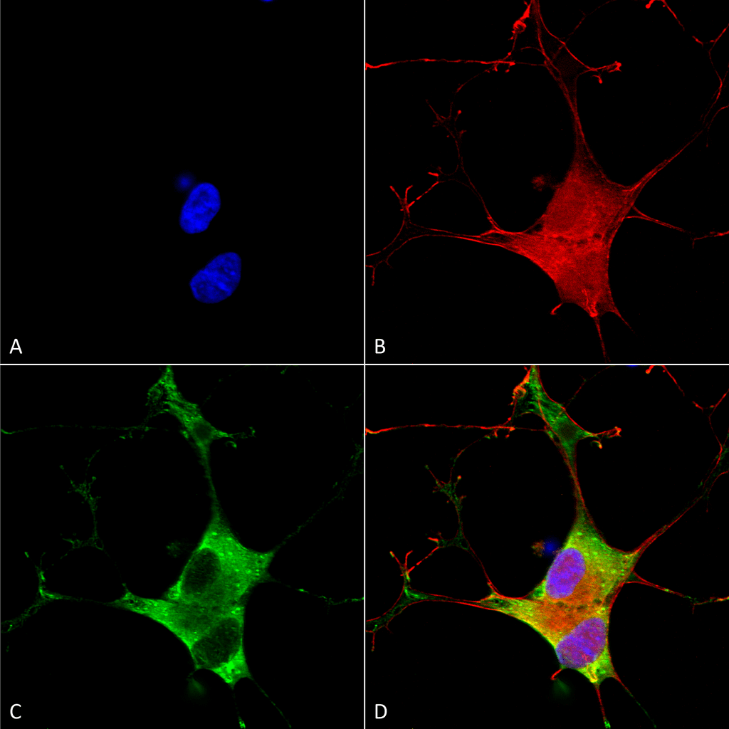 Immunocytochemistry/Immunofluorescence analysis using Mouse Anti-EAAC1 Monoclonal Antibody, Clone S180-41 (56520). Tissue: Neuroblastoma cells (SH-SY5Y). Species: Human. Fixation: 4% PFA for 15 min. Primary Antibody: Mouse Anti-EAAC1 Monoclonal Antibody (56520) at 1:100 for overnight at 4°C with slow rocking. Secondary Antibody: AlexaFluor 488 at 1:1000 for 1 hour at RT. Counterstain: Phalloidin-iFluor 647 (red) F-Actin stain; Hoechst (blue) nuclear stain at 1:800, 1.6mM for 20 min at RT. (A) Hoechst (blue) nuclear stain. (B) Phalloidin-iFluor 647 (red) F-Actin stain. (C) EAAC1 Antibody (D) Composite.
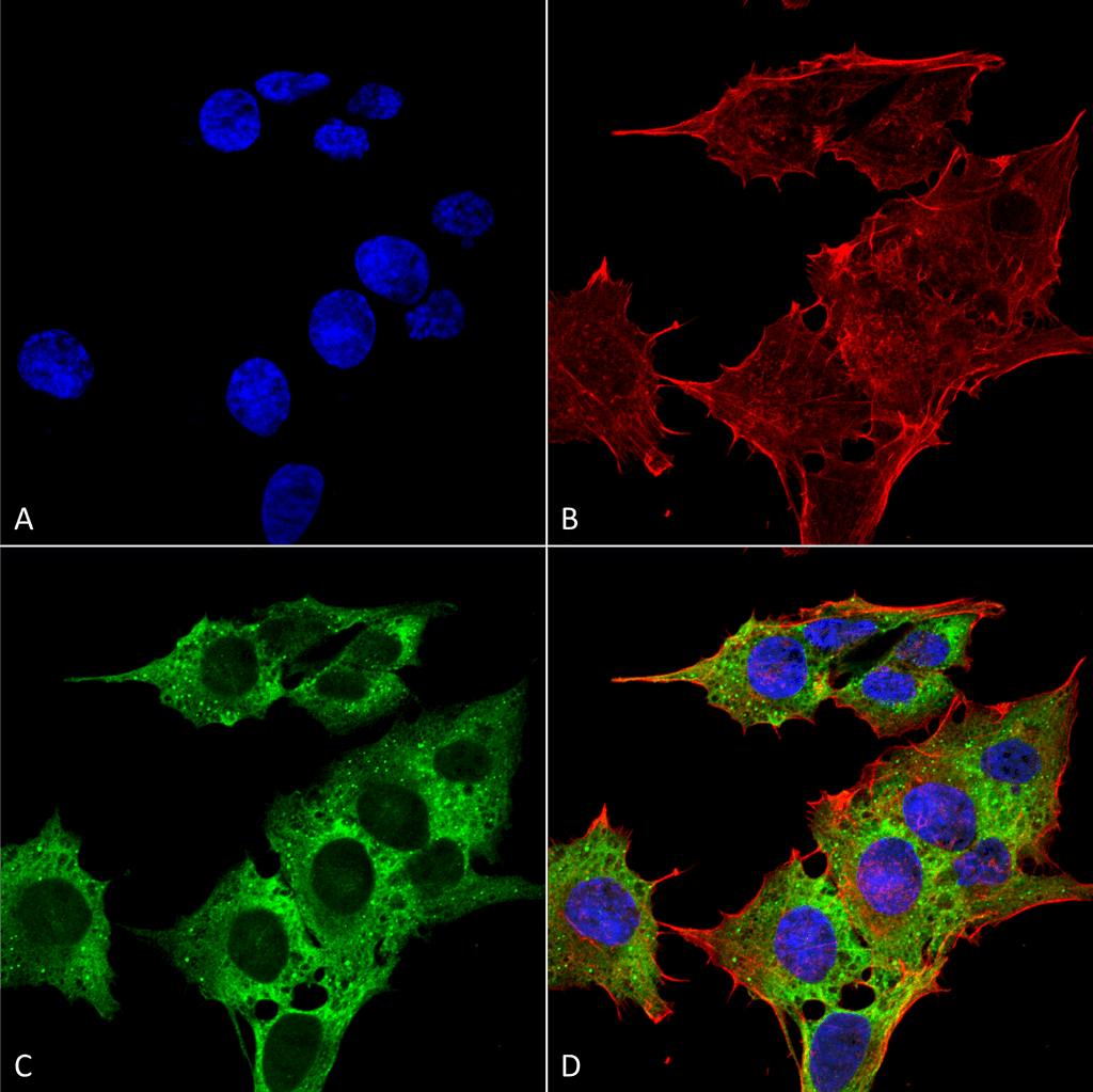 Immunocytochemistry/Immunofluorescence analysis using Mouse Anti-EAAC1 Monoclonal Antibody, Clone S180-41 (56520). Tissue: Neuroblastoma cell line (SK-N-BE). Species: Human. Fixation: 4% Formaldehyde for 15 min at RT. Primary Antibody: Mouse Anti-EAAC1 Monoclonal Antibody (56520) at 1:100 for 60 min at RT. Secondary Antibody: Goat Anti-Mouse ATTO 488 at 1:200 for 60 min at RT. Counterstain: Phalloidin Texas Red F-Actin stain; DAPI (blue) nuclear stain at 1:1000, 1:5000 for 60 min at RT, 5 min at RT. Localization: Cytoplasm, Membrane . Magnification: 60X. (A) DAPI (blue) nuclear stain. (B) Phalloidin Texas Red F-Actin stain. (C) EAAC1 Antibody. (D) Composite.
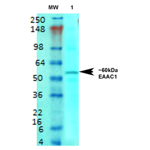 Western Blot analysis of Rat brain membrane lysate showing detection of EAAT3 protein using Mouse Anti-EAAT3 Monoclonal Antibody, Clone S180-41 (56520). Primary Antibody: Mouse Anti-EAAT3 Monoclonal Antibody (56520) at 1:1000.
 - -
- -
Antibody DetailsProduct DetailsReactive Species Human Host Species Mouse Immunogen Fusion protein corresponding to aa 1-524 (full-length) of human EAAC1 (accession no. NP_004161.4). Product Concentration 1.0 mg/ml Formulation PBS, pH 7.4, 50% glycerol, 0.09% sodium azide.Purified by Protein G affinity chromatography. State of Matter Liquid Product Preparation Purified by Protein G affinity chromatography Storage and Handling This product is stable for at least 1 year at -20°C. Freeze in multiple aliquots to avoid repeated freeze-thaw cycles. Regulatory Status For in vitro investigational use only. Not for
use in therapeutic or diagnostic procedures. Country of Origin USA Shipping Next Day 2-8°C Applications and Recommended Usage? Quality Tested by Leinco Immunoblotting: use at 1-5ug/mL. A band of ~60kDa is detected.
These are recommended concentrations. Enduser should determine optimal concentrations for their applications. Positive control: rat brain membranes. Each investigator should determine their own optimal working dilution for specific applications. See directions on lot specific datasheets, as information may periodically change. DescriptionDescriptionSpecificity This antibody recognizes human EAAC1. Background Excitatory amino-acid carrier 1 (EAAC1) is a high-affinity Na+-dependent L- glutamate/D,L-aspartate cell membrane transport protein. It is expressed in brain as well as in several non-nervous system tissues. In brain, EAAC1 is the primary neuronal glutamate transporter. It has a polarized distribution in cells and functions mainly perisynaptically to transport glutamate from the extracellular environment. In the kidney it is involved in renal acidic amino acid reabsorption and amino acid metabolism. EAAC1- associated protein GTRAP3-18 may be important in regulating the metabolic function of EAAC1. Function Sodium-dependent, high-affinity amino acid transporter that mediates the uptake of L-glutamate and also L-aspartate and D-aspartate (PubMed:7914198, PubMed:7521911, PubMed:8857541, PubMed:26690923, PubMed:21123949). Can also transport L-cysteine (PubMed:21123949). Functions as a symporter that transports one amino acid molecule together with two or three Na(+) ions and one proton, in parallel with the counter-transport of one K(+) ion (PubMed:7521911, PubMed:8857541, PubMed:26690923). Mediates Cl(-) flux that is not coupled to amino acid transport; this avoids the accumulation of negative charges due to aspartate and Na(+) symport (PubMed:8857541, PubMed:26690923). Plays an important role in L-glutamate and L-aspartate reabsorption in renal tubuli (PubMed:21123949). Plays a redundant role in the rapid removal of released glutamate from the synaptic cleft, which is essential for terminating the postsynaptic action of glutamate (By similarity). Contributes to glutathione biosynthesis and protection against oxidative stress via its role in L-glutamate and L-cysteine transport (By similarity). Negatively regulated by ARL6IP5 (By similarity). {UniProtKB:P51906, UniProtKB:P51907, PubMed:21123949, PubMed:26690923, PubMed:7521911, PubMed:7914198, PubMed:8857541}. NCBI Gene Bank ID UniProt.org Research Area Neuroscience References & CitationsTechnical Protocols  ICC/IF Certificate of Analysis |
Formats Available
 Products are for research use only. Not for use in diagnostic or therapeutic procedures.