

Anti-Entamoeba Histolytica Library Pack [5 Clones]


Data
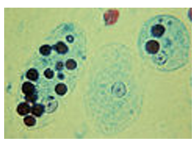 Trophozoites of Entamoeba histolytica with ingested erythrocytes.
 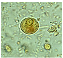 Immature Entamoeba histolytica cyst.
 - -
- -
Antibody DetailsProduct DetailsReactive Species E. histolytica Host Species Mouse Immunogen Entamoeba histolytica HK-9. Product Concentration Lot Specific Formulation This monoclonal antibody is formulated in phosphate buffered saline (PBS) pH 7.2 - 7.4 with no carrier protein or preservatives added. State of Matter Liquid Product Preparation All antibodies in this library pack are manufactured under strict ISO 9001 and ISO 13485 Quality Management Systems at our premier facility in St. Louis, Missouri, USA. We guarantee the lot-to-lot consistency and rigorous quality control required for critical diagnostic and clinical workflows. Storage and Handling These antibodies are stable for at least one (1) year at -20°C to -70°C. Store product in appropriate aliquots to avoid multiple freeze-thaw cycles. Country of Origin USA Shipping Next Day 2-8°C Applications and Recommended Usage? Quality Tested by Leinco These antibodies have been qualified for use in ELISA to detect E. histolytica trophozoites and cysts.
End users should determine optimal concentrations for their applications. Each investigator should determine their own optimal working dilution for specific applications. See directions on lot specific datasheets, as information may periodically change. DescriptionDescriptionSpecificity Target Specificity and Broad reactivity: All of these monoclonal antibodies recognizes both Entamoeba histolytica trophozoites (the active, feeding stage) and cysts (the dormant, infective stage). Library Pack Advantages: By purchasing this library pack, developers can efficiently test multiple paratopes and isotypes in parallel to optimize assay sensitivity and specificity. Included Clones (100 µg each): • Clone EH34.5 (Isotype IgG1) • Clone EH123.5 (Isotype IgG1) • Clone EH147.5 (Isotype IgM) • Clone EH304.6 (Isotype IgG3) • Clone EH357.8 (Isotype IgM) Background Entamoeba histolytica is an anaerobic parasitic protozoan that predominantly infects humans and other primates causing amoebiasis. In the vast majority of cases, infection is asymptomatic and the carrier is unaware they are infected. However, in an estimated 10% of cases E. histolytica causes disease. Once the trophozoites are excysted they colonize the large bowel, remaining on the surface of the mucus layer and feeding on bacteria and food particles. Occasionally, and in response to unknown stimuli, trophozoites move through the mucus layer where they come in contact with the epithelial cell layer and start the pathological process. Ideal for IVD Assay Development Finding the perfect capture and detection combination is critical for diagnostic manufacturing. This library pack provides 5 distinct monoclonal antibody clones (IgG1, IgG3, and IgM isotypes), making it the perfect raw material for matched-pair screening in Sandwich ELISAs, rapid lateral flow tests (RDTs), and other in vitro diagnostic platforms for amoebiasis. Accelerate Vaccine Development For researchers developing vaccines against E. histolytica, these highly specific monoclonal antibodies serve as reliable tools for antigen quantification, immunogenicity assays, and evaluating vaccine efficacy. Generated against the Entamoeba histolytica HK-9 strain, they accurately detect both cysts and trophozoites. Reliable Research Use Only (RUO) Reagents Guarantee reproducibility in your parasitology research. Whether you are studying the pathological processes of amoebiasis, host-parasite interactions, or conducting routine ELISA protocols, this 5-clone library provides comprehensive coverage of E. histolytica life stages. Matched Pair This 5-clone library pack is designed to empower IVD manufacturers to build their own optimized, platform-specific assays. Because antibody behavior shifts depending on your specific matrix, buffer conditions, and platform (e.g., Sandwich ELISA, lateral flow RDTs, or CLIA), we highly recommend conducting a comprehensive matrix screen. Testing all 5 distinct monoclonal clones (incorporating IgG1, IgG3, and IgM isotypes) as both capture and detection reagents allows you to empirically identify the absolute highest-affinity, lowest-background combination for your unique diagnostic layout.. By evaluating all five clones simultaneously, you ensure that the chosen pair is fully optimized for your specific surface chemistry—whether it is a polystyrene ELISA plate, nitrocellulose membrane, or magnetic bead. Research Area Infectious Disease References & Citations1.) Cooney J, Siakavellas SI, Chiodini PL, Mahadeva U, Godbole G, Pollok RC, Smith PJ. Recent advances in the diagnosis and management of amoebiasis. Frontline Gastroenterol. 2024 Oct 7;16(1):e102554. doi: 10.1136/flgastro-2023-102554. Erratum in: Frontline Gastroenterol. 2025 Apr 8;16(3):e2. doi: 10.1136/flgastro-2023-102554corr1. PMID: 41809248; PMCID: PMC12969968. 2.) Tanyuksel M, Petri WA Jr. Laboratory diagnosis of amebiasis. Clin Microbiol Rev. 2003 Oct;16(4):713-29. doi: 10.1128/CMR.16.4.713-729.2003. PMID: 14557296; PMCID: PMC207118. Technical Protocols Immunoassays Certificate of Analysis |
Related Products
- -
- -
 Products are for research use only. Not for use in diagnostic or therapeutic procedures.