

Anti-GABA(B)R2 Antibody (56523)


Anti-GABA(B)R2 Antibody (56523)
Product No.: 56523
- -
- -
Clone S81-2 Target GABA(B)R2 Formats AvailableView All Product Type Monoclonal Alternate Names GABA-B receptor 2, GABA-B-R2, GABA-BR2, GABABR2, Gb2, G-protein coupled receptor 51 Isotype Mouse IgG1 Applications IHC , WB , ICC/IF |
Data
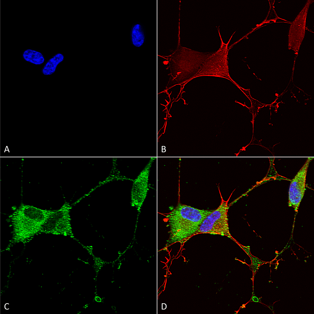 Immunocytochemistry/Immunofluorescence analysis using Mouse Anti-GABA-B Receptor 2 Monoclonal Antibody, Clone S81-2 (56523). Tissue: Neuroblastoma cells (SH-SY5Y). Species: Human. Fixation: 4% PFA for 15 min. Primary Antibody: Mouse Anti-GABA-B Receptor 2 Monoclonal Antibody (56523) at 1:100 for overnight at 4°C with slow rocking. Secondary Antibody: AlexaFluor 488 at 1:1000 for 1 hour at RT. Counterstain: Phalloidin-iFluor 647 (red) F-Actin stain; Hoechst (blue) nuclear stain at 1:800, 1.6mM for 20 min at RT. (A) Hoechst (blue) nuclear stain. (B) Phalloidin-iFluor 647 (red) F-Actin stain. (C) GABA-B Receptor 2 Antibody (D) Composite.
 Western Blot analysis of Rat Brain Membrane showing detection of ~105 kDa GABA B Receptor 2 protein using Mouse Anti-GABA B Receptor 2 Monoclonal Antibody, Clone S81-2 (56523). Lane 1: MW Ladder. Lane 2: Rat Brain Membrane (10 µg). . Load: 10 µg. Block: 5% milk. Primary Antibody: Mouse Anti-GABA B Receptor 2 Monoclonal Antibody (56523) at 1:1000 for 1 hour at RT. Secondary Antibody: Goat Anti-Mouse IgG: HRP at 1:200 for 1 hour at RT. Color Development: TMB solution for 10 min at RT. Predicted/Observed Size: ~105 kDa.
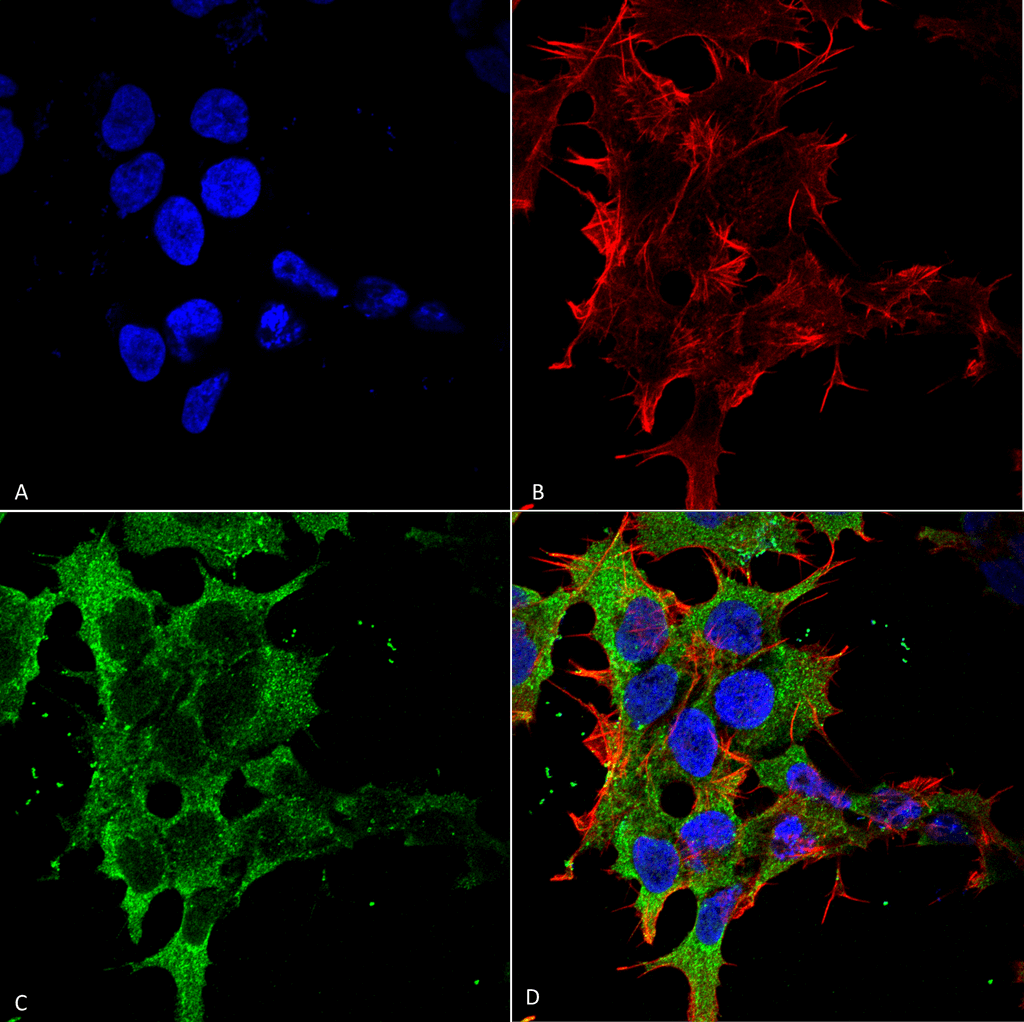 Immunocytochemistry/Immunofluorescence analysis using Mouse Anti-GABA-B Receptor 2 Monoclonal Antibody, Clone S81-2 (56523). Tissue: Neuroblastoma cell line (SK-N-BE). Species: Human. Fixation: 4% Formaldehyde for 15 min at RT. Primary Antibody: Mouse Anti-GABA-B Receptor 2 Monoclonal Antibody (56523) at 1:100 for 60 min at RT. Secondary Antibody: Goat Anti-Mouse ATTO 488 at 1:100 for 60 min at RT. Counterstain: Phalloidin Texas Red F-Actin stain; DAPI (blue) nuclear stain at 1:1000, 1:5000 for 60min RT, 5min RT. Localization: Cell Membrane. Magnification: 60X. (A) DAPI (blue) nuclear stain. (B) Phalloidin Texas Red F-Actin stain. (C) GABA-B Receptor 2 Antibody. (D) Composite.
 - -
- -
Antibody DetailsProduct DetailsReactive Species Human ⋅ Mouse ⋅ Rat Host Species Mouse Immunogen Fusion protein corresponding to aa 861-912 of rat GABA(B)R2 (accession no. NP_113990.1). Product Concentration 1.0 mg/ml Formulation PBS, pH 7.4, 50% glycerol, 0.09% sodium azide.Purified by Protein G affinity chromatography. State of Matter Liquid Product Preparation Purified by Protein G affinity chromatography Storage and Handling This product is stable for at least 1 year at -20°C. Freeze in multiple aliquots to avoid repeated freeze-thaw cycles. Regulatory Status For in vitro investigational use only. Not for
use in therapeutic or diagnostic procedures. Country of Origin USA Shipping Next Day 2-8°C Applications and Recommended Usage? Quality Tested by Leinco Immunoblotting: use at 1-2ug/mL. A band of ~105kDa is detected.
Immunohistochemistry: use at 1-5ug/mL. These are recommended concentrations. Enduser should determine optimal concentrations for their applications. Positive control: Rat brain membranes. Each investigator should determine their own optimal working dilution for specific applications. See directions on lot specific datasheets, as information may periodically change. DescriptionDescriptionSpecificity This antibody recognizes human, mouse,
and rat GABA(B)R2. It does not cross-react with GABA(B)R1. Background GABA (g-aminobutyric acid) is the primary inhibitory neurotransmitter in the central nervous system and interacts with three different receptors: GABA(A), GABA(B), and GABA(C). GABA(B) receptor is coupled to G proteins that modulate slow inhibitory synaptic transmission. Functional GABA(B) receptors form heterodimers of GABA(B)R1 and GABA(B)R2 in which GABA(B)R1 binds a ligand and GABA(B)R2 is the primary G protein contact site. Function Component of a heterodimeric G-protein coupled receptor for GABA, formed by GABBR1 and GABBR2 (PubMed:9872315, PubMed:9872317, PubMed:9872744). Within the heterodimeric GABA receptor, only GABBR1 seems to bind agonists, while GABBR2 mediates coupling to G proteins (PubMed:9872317, PubMed:10658574). Ligand binding causes a conformation change that triggers signaling via guanine nucleotide-binding proteins (G proteins) and modulates the activity of down-stream effectors, such as adenylate cyclase (PubMed:9872315, PubMed:9872317, Ref.4, PubMed:10075644, PubMed:9872744, PubMed:10924501). Signaling inhibits adenylate cyclase, stimulates phospholipase A2, activates potassium channels, inactivates voltage-dependent calcium-channels and modulates inositol phospholipid hydrolysis (PubMed:9872315, PubMed:9872317, PubMed:10457184, PubMed:9872744, PubMed:10924501). Plays a critical role in the fine-tuning of inhibitory synaptic transmission (PubMed:9872317, PubMed:10457184, PubMed:9872744). Pre-synaptic GABA receptor inhibits neurotransmitter release by down-regulating high-voltage activated calcium channels, whereas postsynaptic GABA receptor decreases neuronal excitability by activating a prominent inwardly rectifying potassium (Kir) conductance that underlies the late inhibitory postsynaptic potentials (PubMed:9872744, PubMed:10924501). Not only implicated in synaptic inhibition but also in hippocampal long-term potentiation, slow wave sleep, muscle relaxation and antinociception (By similarity). {UniProtKB:O75899, PubMed:10075644, PubMed:10457184, PubMed:10658574, PubMed:10924501, PubMed:9872315, PubMed:9872317, PubMed:9872744, Ref.4}. NCBI Gene Bank ID UniProt.org Research Area Neuroscience References & CitationsTechnical Protocols  ICC/IF Certificate of Analysis |
Formats Available
 Products are for research use only. Not for use in diagnostic or therapeutic procedures.