


Data
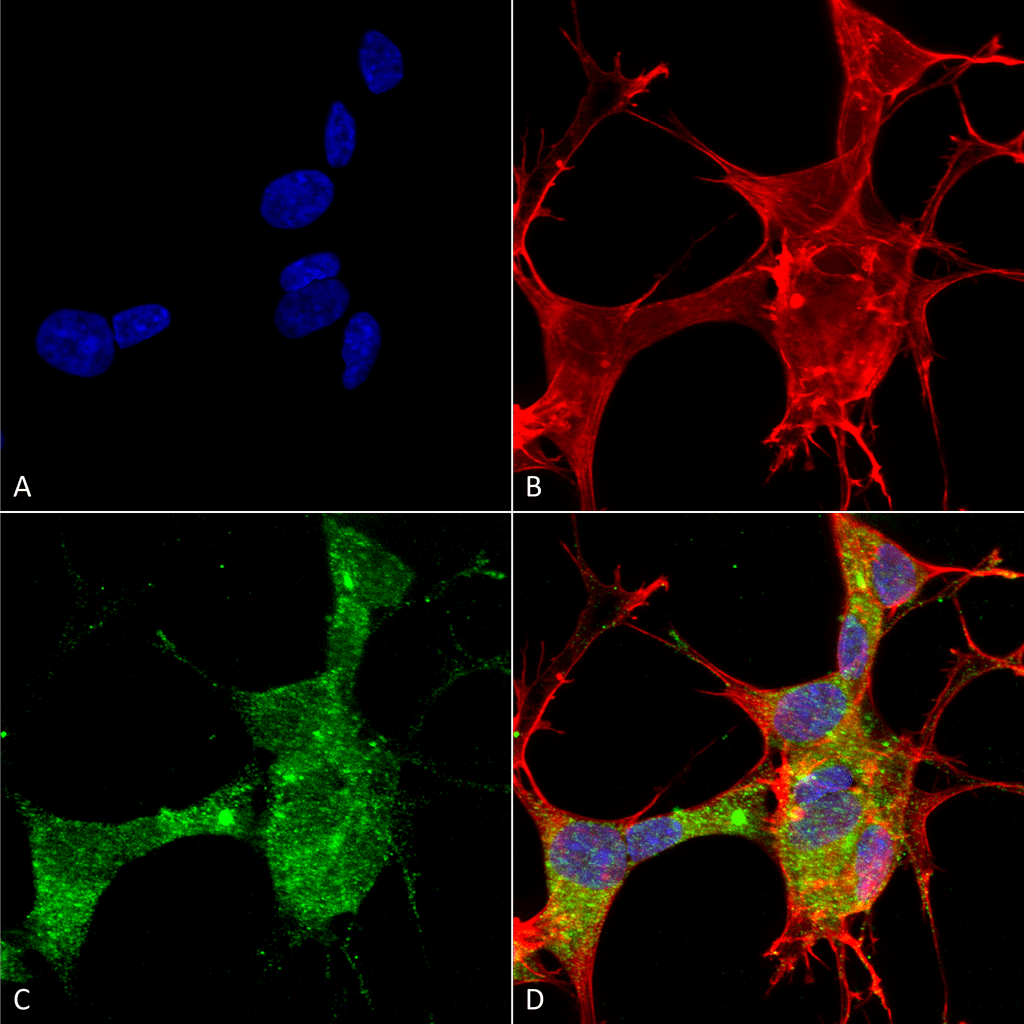 Immunocytochemistry/Immunofluorescence analysis using Mouse Anti-GFAP Monoclonal Antibody, Clone S206A-8 (56566). Tissue: Neuroblastoma cells (SH-SY5Y). Species: Human. Fixation: 4% PFA for 15 min. Primary Antibody: Mouse Anti-GFAP Monoclonal Antibody (56566) at 1:50 for overnight at 4°C with slow rocking. Secondary Antibody: AlexaFluor 488 at 1:1000 for 1 hour at RT. Counterstain: Phalloidin-iFluor 647 (red) F-Actin stain; Hoechst (blue) nuclear stain at 1:800, 1.6mM for 20 min at RT. (A) Hoechst (blue) nuclear stain. (B) Phalloidin-iFluor 647 (red) F-Actin stain. (C) GFAP Antibody (D) Composite.
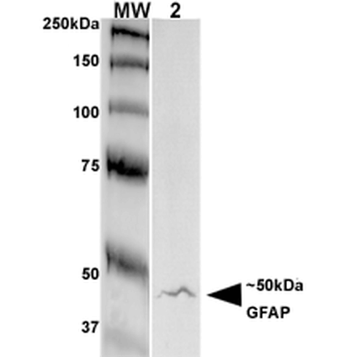 Western Blot analysis of Rat Brain Membrane showing detection of GFAP protein using Mouse Anti-GFAP Monoclonal Antibody, Clone S206A-8 (56566). Primary Antibody: Mouse Anti-GFAP Monoclonal Antibody (56566) at 1:250.
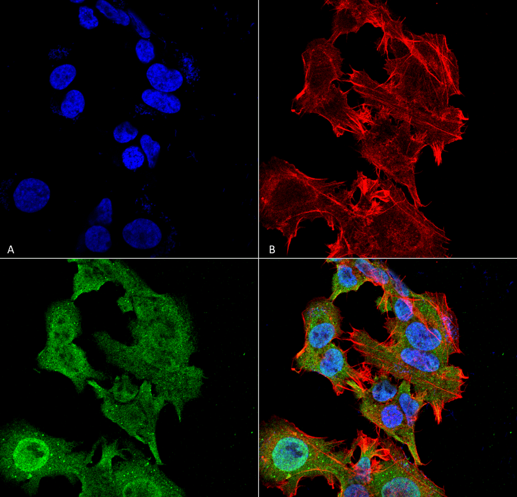 Immunocytochemistry/Immunofluorescence analysis using Mouse Anti-GFAP Monoclonal Antibody, Clone S206A-8 (56566). Tissue: Neuroblastoma cell line (SK-N-BE). Species: Human. Fixation: 4% Formaldehyde for 15 min at RT. Primary Antibody: Mouse Anti-GFAP Monoclonal Antibody (56566) at 1:100 for 60 min at RT. Secondary Antibody: Goat Anti-Mouse ATTO 488 at 1:100 for 60 min at RT. Counterstain: Phalloidin Texas Red F-Actin stain; DAPI (blue) nuclear stain at 1:1000, 1:5000 for 60min RT, 5min RT. Localization: Cytoplasm . Magnification: 60X. (A) DAPI (blue) nuclear stain. (B) Phalloidin Texas Red F-Actin stain. (C) GFAP Antibody. (D) Composite.
 - -
- -
Antibody DetailsProduct DetailsReactive Species Human ⋅ Mouse ⋅ Rat Host Species Mouse Immunogen Synthetic peptide corresponding to aa 411-422 (KTVEMRDGEVIK) of human GFAP. This sequence is 100 Product Concentration 1.0 mg/ml Formulation PBS, pH 7.4, 0.1% sodium azide, 50% glycerol. State of Matter Liquid Product Preparation Purified by Protein G affinity chromatography Storage and Handling This product is stable for at least one (1) year at -20°C. Regulatory Status For in vitro investigational use only. Not intended for therapeutic or diagnostic procedures. Country of Origin USA Shipping Next Day 2-8°C Applications and Recommended Usage? Quality Tested by Leinco Immunoblotting: use at 1-4ug/mL. A band of ~50kDa is detected.
Immunofluorescence: use at 10ug/mL. These are recommended concentrations. Endusers should determine optimal concentrations for their application. Each investigator should determine their own optimal working dilution for specific applications. See directions on lot specific datasheets, as information may periodically change. DescriptionDescriptionSpecificity This antibody recognizes human, mouse and rat GFAP. It cross-reacts with GFAPR416W and other GFAP mutant proteins. Background Glial fibrillary acidic protein (GFAP) is a major structural component of astrocytes. Phosphorylation of GFAP, and its association with annexin II-p2 and S-100, regulates GFAP polymerization. One of the first events that occurs during astrocyte proliferation is increased GFAP expression. Antibodies to GFAP have been detected in individuals with dementia, although the significance of this has not been conclusively determined. Function GFAP, a class-III intermediate filament, is a cell-specific marker that, during the development of the central nervous system, distinguishes astrocytes from other glial cells. NCBI Gene Bank ID UniProt.org Research Area Neuroscience References & CitationsTechnical Protocols  ICC/IF Certificate of Analysis |
