

Anti-Glial Fibrillary Acidic Protein (GFAP) Antibody (56575)

Anti-Glial Fibrillary Acidic Protein (GFAP) Antibody (56575)
Product No.: 56575
- -
- -
Clone S206B-9 Target Glial Fibrillary Acidic Protein (GFAP) Formats AvailableView All Product Type Monoclonal Alternate Names GFAP Isotype Mouse IgG1 Applications ICC , IF , IHC , WB |
Data
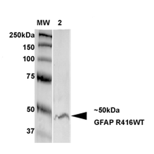 Western Blot analysis of Rat Brain Membrane showing detection of GFAP protein using Mouse Anti-GFAP Monoclonal Antibody, Clone S206B-9 (56575). Primary Antibody: Mouse Anti-GFAP Monoclonal Antibody (56575) at 1:250.
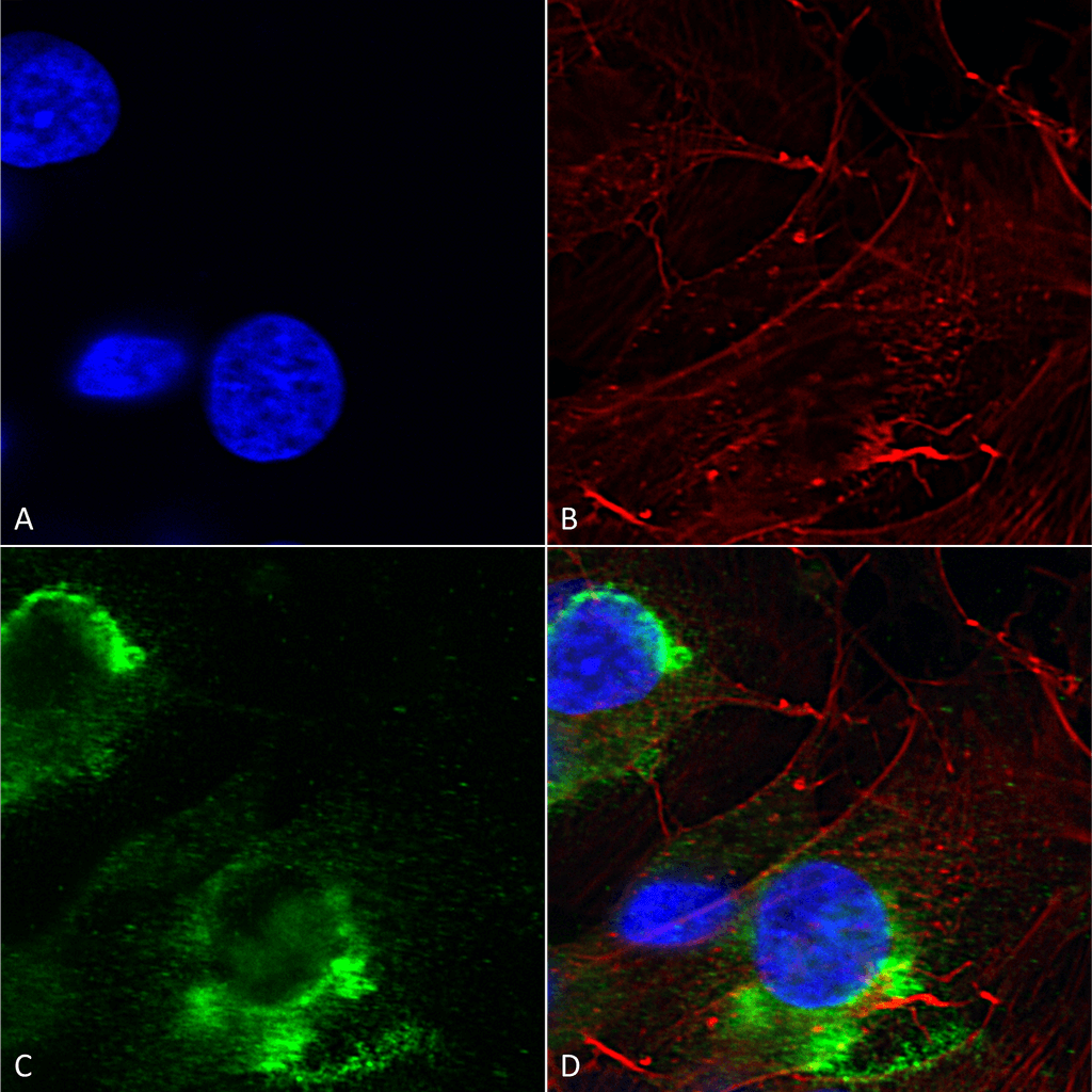 Immunocytochemistry/Immunofluorescence analysis using Mouse Anti-GFAP R416WT Monoclonal Antibody, Clone S206B-9 (56575). Tissue: Neuroblastoma cells (SH-SY5Y). Species: Human. Fixation: 4% PFA for 15 min. Primary Antibody: Mouse Anti-GFAP R416WT Monoclonal Antibody (56575) at 1:200 for overnight at 4°C with slow rocking. Secondary Antibody: AlexaFluor 488 at 1:1000 for 1 hour at RT. Counterstain: Phalloidin-iFluor 647 (red) F-Actin stain; Hoechst (blue) nuclear stain at 1:800, 1.6mM for 20 min at RT. (A) Hoechst (blue) nuclear stain. (B) Phalloidin-iFluor 647 (red) F-Actin stain. (C) GFAP R416WT Antibody (D) Composite.
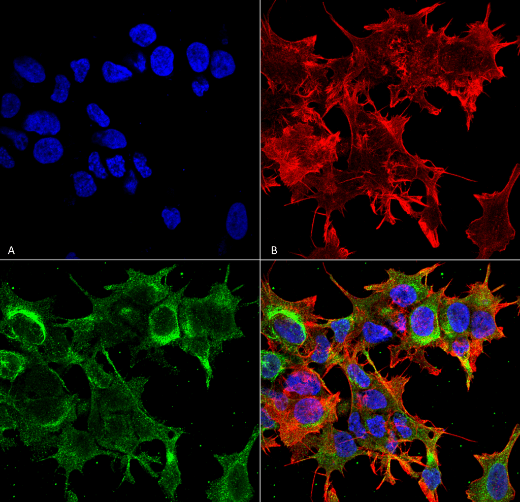 Immunocytochemistry/Immunofluorescence analysis using Mouse Anti-GFAP R416WT Monoclonal Antibody, Clone S206B-9 (56575). Tissue: Neuroblastoma cell line (SK-N-BE). Species: Human. Fixation: 4% Formaldehyde for 15 min at RT. Primary Antibody: Mouse Anti-GFAP R416WT Monoclonal Antibody (56575) at 1:100 for 60 min at RT. Secondary Antibody: Goat Anti-Mouse ATTO 488 at 1:100 for 60 min at RT. Counterstain: Phalloidin Texas Red F-Actin stain; DAPI (blue) nuclear stain at 1:1000, 1:5000 for 60min RT, 5min RT. Localization: Cytoplasm . Magnification: 60X. (A) DAPI (blue) nuclear stain. (B) Phalloidin Texas Red F-Actin stain. (C) GFAP R416WT Antibody. (D) Composite.
 - -
- -
Antibody DetailsProduct DetailsReactive Species Human ⋅ Mouse ⋅ Rat Host Species Mouse Immunogen Synthetic peptide corresponding to aa 411-422 (KTVEMRDGEVIK) of human GFAP. This sequence is 100 Product Concentration 1.0 mg/ml Formulation PBS, pH 7.4, 0.1% sodium azide, 50% glycerol. State of Matter Liquid Product Preparation Purified by Protein G affinity chromatography Storage and Handling This product is stable for at least one (1) year at -20°C. Regulatory Status For in vitro investigational use only. Not intended for therapeutic or diagnostic procedures. Country of Origin USA Shipping Next Day 2-8°C Applications and Recommended Usage? Quality Tested by Leinco Immunoblotting: use at 1-5ug/mL. A band of ~50kDa is detected.
Immunofluorescence: use at 10ug/mL. These are recommended concentrations. Endusers should determine optimal concentrations for their application. Each investigator should determine their own optimal working dilution for specific applications. See directions on lot specific datasheets, as information may periodically change. DescriptionDescriptionSpecificity This antibody recognizes human, mouse, and rat GFAP. Background Glial fibrillary acidic protein (GFAP) is an intermediate filament protein that is expressed by numerous cell types of the central nervous system. It is involved in many important CNS processes, including cell communication and the functioning of the blood brain barrier. It is closely related to its non-epithelial family members, vimentin, desmin, and peripherin, which are all involved in the structure and function of the cell's cytoskeleton. GFAP is thought to help to maintain astrocyte strength and shape. There are multiple disorders associated with improper GFAP regulation. GFAP levels are used as a marker of neurologic damage in adults who suffer strokes and traumatic brain injuries. Function GFAP, a class-III intermediate filament, is a cell-specific marker that, during the development of the central nervous system, distinguishes astrocytes from other glial cells. NCBI Gene Bank ID UniProt.org Research Area Neuroscience References & CitationsTechnical ProtocolsICC IF   Certificate of Analysis |
