

Anti-GluA1-GluR1 Antibody (56547)

Anti-GluA1-GluR1 Antibody (56547)
Product No.: 56547
- -
- -
Clone S355-1 Target GluA1-GluR1 Formats AvailableView All Product Type Monoclonal Alternate Names GluR-1, AMPA-selective glutamate receptor 1, GluR-A, GluR-K1, Glutamate receptor ionotropic, AMPA 1, GluA1 Isotype Mouse IgG1 Applications IHC , WB , ICC/IF |
Data
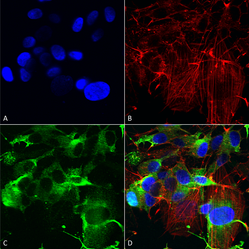 Immunocytochemistry/Immunofluorescence analysis using Mouse Anti-GluA1/GluR1 Monoclonal Antibody, Clone S355-1 (56547). Tissue: Neuroblastoma cells (SH-SY5Y). Species: Human. Fixation: 4% PFA for 15 min. Primary Antibody: Mouse Anti-GluA1/GluR1 Monoclonal Antibody (56547) at 1:200 for overnight at 4°C with slow rocking. Secondary Antibody: AlexaFluor 488 at 1:1000 for 1 hour at RT. Counterstain: Phalloidin-iFluor 647 (red) F-Actin stain; Hoechst (blue) nuclear stain at 1:800, 1.6mM for 20 min at RT. (A) Hoechst (blue) nuclear stain. (B) Phalloidin-iFluor 647 (red) F-Actin stain. (C) GluA1/GluR1 Antibody (D) Composite.
 Western Blot analysis of Rat Brain Membrane showing detection of ~100 kDa GluA1-GluR1 protein using Mouse Anti-GluA1-GluR1 Monoclonal Antibody, Clone S355-1 (56547). Load: 10 µg. Block: 5% milk + TBST. Primary Antibody: Mouse Anti-GluA1-GluR1 Monoclonal Antibody (56547) at 1:2000 for 1 hour at RT. Secondary Antibody: Goat Anti-Mouse HRP at 1:200 for 1 hour at RT. Predicted/Observed Size: ~100 kDa.
 Immunocytochemistry/Immunofluorescence analysis using Mouse Anti-GluA1/GluR1 Glutamate Receptor Monoclonal Antibody, Clone S355-1 (56547). Tissue: Neuroblastoma cell line (SK-N-BE). Species: Human. Fixation: 4% Formaldehyde for 15 min at RT. Primary Antibody: Mouse Anti-GluA1/GluR1 Glutamate Receptor Monoclonal Antibody (56547) at 1:100 for 60 min at RT. Secondary Antibody: Goat Anti-Mouse ATTO 488 at 1:100 for 60 min at RT. Counterstain: Phalloidin Texas Red F-Actin stain; DAPI (blue) nuclear stain at 1:1000, 1:5000 for 60min RT, 5min RT. Localization: Cell Membrane, Cell Junction. Magnification: 60X. (A) DAPI (blue) nuclear stain. (B) Phalloidin Texas Red F-Actin stain. (C) GluA1/GluR1 Glutamate Receptor Antibody. (D) Composite.
 - -
- -
Antibody DetailsProduct DetailsReactive Species Mouse ⋅ Rat Host Species Mouse Immunogen Fusion protein corresponding to aa 1-389 (extracellular N-terminus) of rat GluA1/GluR1 (accession no. P19490). Product Concentration Lot Specific Formulation PBS, pH 7.4; 50% glycerol, 0.09% sodium azide. State of Matter Liquid Product Preparation Purified by Protein G affinity chromatography Storage and Handling This antibody is stable for at least one (1) year at -20°C. Avoid repeated freezing
and thawing. Regulatory Status For in vitro investigational use only. Not for
use in therapeutic or diagnostic procedures. Country of Origin USA Shipping Next Day 2-8°C Applications and Recommended Usage? Quality Tested by Leinco Immunoblotting: use at 1ug/mL. A band of ~100kDa is detected.
Positive control: Rat brain lysate. These are recommended concentrations. User should determine optimal concentrations for their application. Each investigator should determine their own optimal working dilution for specific applications. See directions on lot specific datasheets, as information may periodically change. DescriptionDescriptionSpecificity This antibody recognizes mouse and rat
GluA1/GluR1. It does not cross-react with
GluR2. Background Regulation of alpha-amino-3-hydroxy-5- methyl-4-isoxazole propionic acid receptors (AMPARs) plays a key role in altering excitatory synaptic transmission in the CNS. Interestingly, the regulatory mechanisms differ between distinct subunits of AMPAR, which range from glutamate receptor 1 (GluR1) to GluR4 (also referred to as GluA1–4). For example, subunits with a long intracellular carboxy terminus (i.e., GluR1, GluR2L, and GluR4) are involved in activity- dependent synaptic targeting of AMPAR, whereas those with a shorter carboxy terminus (i.e., GluR2, GluR3, and GluR4s) seem to maintain basal synaptic transmission. GluR1 has several phosphorylation sites on the intracellular carboxy terminus. Many of these sites have been demonstrated to play a role in synaptic AMPAR regulation and synaptic plasticity. Function Ionotropic glutamate receptor. L-glutamate acts as an excitatory neurotransmitter at many synapses in the central nervous system. Binding of the excitatory neurotransmitter L-glutamate induces a conformation change, leading to the opening of the cation channel, and thereby converts the chemical signal to an electrical impulse. The receptor then desensitizes rapidly and enters a transient inactive state, characterized by the presence of bound agonist. In the presence of CACNG4 or CACNG7 or CACNG8, shows resensitization which is characterized by a delayed accumulation of current flux upon continued application of glutamate. {PubMed:16793768, PubMed:19265014}. NCBI Gene Bank ID UniProt.org Research Area Neuroscience References & CitationsTechnical Protocols  ICC/IF Certificate of Analysis |
