

Anti-HSP60 [Clone LK-2]


Anti-HSP60 [Clone LK-2]
Product No.: 11101
- -
- -
Clone LK-2 Target Hsp60 Formats AvailableView All Product Type Monoclonal Alternate Names EC 5.6.1.7, 60 kDa chaperonin, Chaperonin 60, CPN60, Heat shock protein 60, HSP-60, Hsp60, HuCHA60, Mitochondrial matrix protein P1, P60 lymphocyte protein Isotype Mouse IgG1 Applications IHC , WB , FCM |
Data
 Immunohistochemistry analysis using Mouse Anti-Hsp60 Monoclonal Antibody, Clone LK-2 (11101). Tissue: colon carcinoma. Species: Human. Fixation: Formalin. Primary Antibody: Mouse Anti-Hsp60 Monoclonal Antibody (11101) at 1:100000 for 12 hours at 4°C. Secondary Antibody: Biotin Goat Anti-Mouse at 1:2000 for 1 hour at RT. Counterstain: Mayer Hematoxylin (purple/blue) nuclear stain at 200 µl for 2 minutes at RT. Localization: Inflammatory cells. Magnification: 40x. This image was produced using an amplifying IHC wash buffer. The antibody has therefore been diluted more than is recommended for other applications.
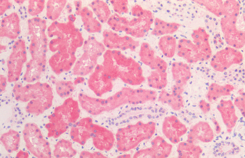 Immunohistochemistry analysis using Mouse Anti-HSP60 Monoclonal Antibody, Clone LK2 (11101). Tissue: Kidney. Species: Human. Fixation: Formalin Fixed, Paraffin Embedded. Primary Antibody: Mouse Anti-HSP60 Monoclonal Antibody (11101).
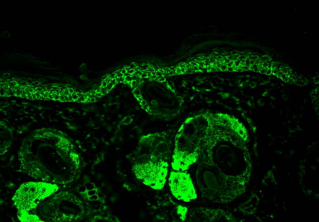 Immunohistochemistry analysis using Mouse Anti-Hsp60 Monoclonal Antibody, Clone LK-2 (11101). Tissue: backskin. Species: Mouse. Fixation: Bouin’s Fixative and paraffin-embedded. Primary Antibody: Mouse Anti-Hsp60 Monoclonal Antibody (11101) at 1:100 for 1 hour at RT. Secondary Antibody: FITC Goat Anti-Mouse (green) at 1:50 for 1 hour at RT. Localization: Cytoplasmic Staining.
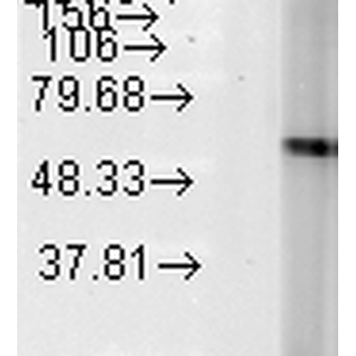 Western Blot analysis of Human Heat Shocked cervical cancer cell line (HeLa) lysate showing detection of Hsp60 protein using Mouse Anti-Hsp60 Monoclonal Antibody, Clone LK-2 (11101). Load: 15 µg. Block: 1.5% BSA for 30 minutes at RT. Primary Antibody: Mouse Anti-Hsp60 Monoclonal Antibody (11101) at 1:1000 for 2 hours at RT. Secondary Antibody: Sheep Anti-Mouse IgG: HRP for 1 hour at RT.
 - -
- -
Antibody DetailsProduct DetailsReactive Species Bacteria ⋅ Bovine ⋅ Canine ⋅ Chicken ⋅ Guinea Pig ⋅ Hamster ⋅ Human ⋅ Monkey ⋅ Mouse ⋅ Porcine ⋅ Rabbit ⋅ Rat ⋅ White Fly ⋅ Yeast Host Species Mouse Immunogen Recombinant human Hsp60 expressed in E. coli. Product Concentration Lot Specific Formulation PBS, pH 7.4. State of Matter Liquid Product Preparation Purified by Protein G affinity chromatography Storage and Handling This antibody is stable for at least one (1) year at -20°C. Avoid repeated freezing and
thawing. Regulatory Status For in vitro investigational use only. Not for use in therapeutic or diagnostic procedures. Country of Origin USA Shipping Next Day 2-8°C Applications and Recommended Usage? Quality Tested by Leinco Immunoblotting: use at 0.25-1ug/mL. A band of ~60 kDa is detected.
Immunohistochemistry: use at 5ug/mLFlow cytometry: use at 10ug/mL These are recommended concentrations. User should determine optimal concentrations for their application. Positive control: Heat-shocked HeLa cell lysate. Each investigator should determine their own optimal working dilution for specific applications. See directions on lot specific datasheets, as information may periodically change. DescriptionDescriptionSpecificity This antibody recognizes human, mouse,
rat, rabbit, bovine, canine, porcine, guinea
pig, hamster, chicken, monkey, white fly,
yeast and bacterial* Hsp60 (60 kDa). The
epitope recognized is within aa 383-419 of
human Hsp60.
*Borellia, E. coli, Helicobacter pylori, M.
bovis, Salmonella typhimurium,
Streptococcus pyogenes, Treponema
hyodysenteriae, Treponema innocense,
Trichinella spiralis, Yersinia enterocolitica. Background Hsp60 is an abundant protein synthesized constitutively in various cell types that is induced to higher concentrations after cell shock. It is present in mitochondria of many mammalian species and has highly similar counterparts in bacteria and plants (where it is localized to chloroplasts). In general, Hsp60 proteins are present in high concentrations, are induced in response to environmental stresses (such as heat shock) are homo-oligomeric structures of 7 or 14 subunits that dissociate reversibly in the presence of Mg2+ and ATP, have ATPase activity, and play a role in folding and assembly of oligomeric protein structures. Hsp60 has been linked to Alzheimer's disease, coronary artery disease, multiple sclerosis, diabetes, and other autoimmune diseases. Function Chaperonin implicated in mitochondrial protein import and macromolecular assembly. Together with Hsp10, facilitates the correct folding of imported proteins. May also prevent misfolding and promote the refolding and proper assembly of unfolded polypeptides generated under stress conditions in the mitochondrial matrix (PubMed:1346131, PubMed:11422376). The functional units of these chaperonins consist of heptameric rings of the large subunit Hsp60, which function as a back-to-back double ring. In a cyclic reaction, Hsp60 ring complexes bind one unfolded substrate protein per ring, followed by the binding of ATP and association with 2 heptameric rings of the co-chaperonin Hsp10. This leads to sequestration of the substrate protein in the inner cavity of Hsp60 where, for a certain period of time, it can fold undisturbed by other cell components. Synchronous hydrolysis of ATP in all Hsp60 subunits results in the dissociation of the chaperonin rings and the release of ADP and the folded substrate protein (Probable). {PubMed:11422376, PubMed:1346131, PubMed:25918392}. NCBI Gene Bank ID UniProt.org Research Area Cell Biology . Immunology . Innate Immunity . Neuroinflammation . Neuroscience . Neuroscience Cell Markers . Heat Shock & Stress Proteins . Mitochondrial Function References & CitationsTechnical Protocols  FCM Certificate of Analysis |
Formats Available
 Products are for research use only. Not for use in diagnostic or therapeutic procedures.