

Anti-LRRK2/Dardarin Antibody (56563)

Anti-LRRK2/Dardarin Antibody (56563)
Product No.: 56563
- -
- -
Clone S138-6 Target LRRK2/Dardarin Formats AvailableView All Product Type Monoclonal Alternate Names EC 2.7.11.1, EC 3.6.5.-, Dardarin Isotype Mouse IgG1 Applications ICC , IF , WB |
Data
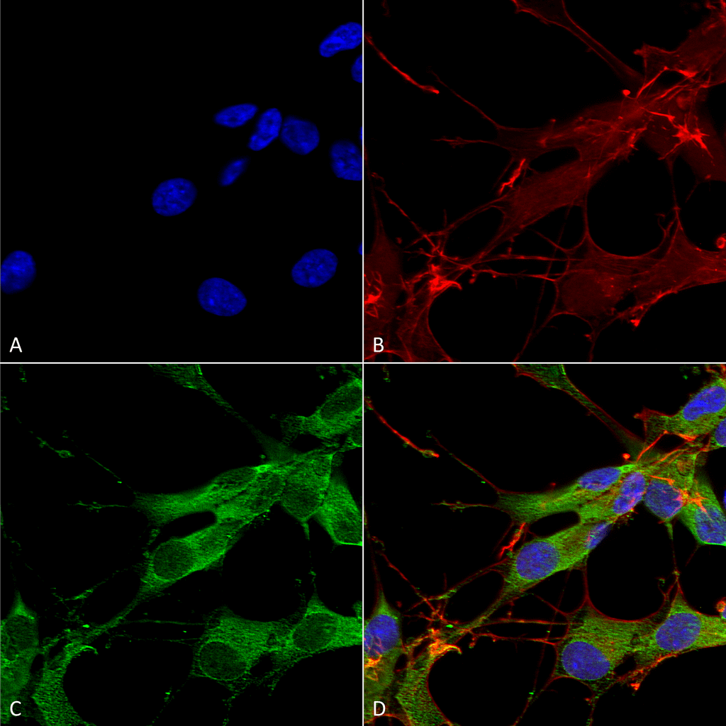 Immunocytochemistry/Immunofluorescence analysis using Mouse Anti-LRRK2/Dardarin Monoclonal Antibody, Clone S138-6 (56563). Tissue: Neuroblastoma cells (SH-SY5Y). Species: Human. Fixation: 4% PFA for 15 min. Primary Antibody: Mouse Anti-LRRK2/Dardarin Monoclonal Antibody (56563) at 1:100 for overnight at 4°C with slow rocking. Secondary Antibody: AlexaFluor 488 at 1:1000 for 1 hour at RT. Counterstain: Phalloidin-iFluor 647 (red) F-Actin stain; Hoechst (blue) nuclear stain at 1:800, 1.6mM for 20 min at RT. (A) Hoechst (blue) nuclear stain. (B) Phalloidin-iFluor 647 (red) F-Actin stain. (C) LRRK2/Dardarin Antibody (D) Composite.
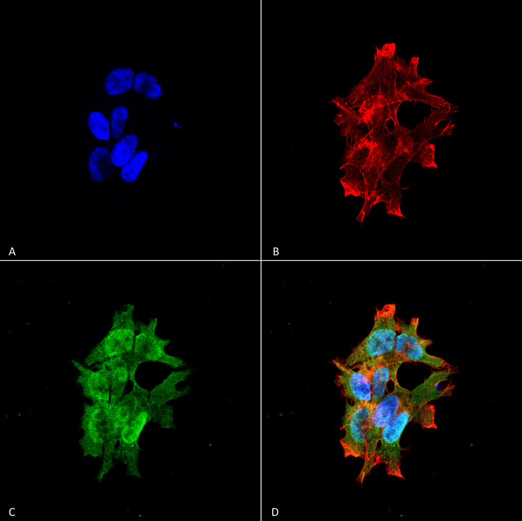 Immunocytochemistry/Immunofluorescence analysis using Mouse Anti-LRRK2/Dardarin Monoclonal Antibody, Clone S138-6 (56563). Tissue: Neuroblastoma cell line (SK-N-BE). Species: Human. Fixation: 4% Formaldehyde for 15 min at RT. Primary Antibody: Mouse Anti-LRRK2/Dardarin Monoclonal Antibody (56563) at 1:100 for 60 min at RT. Secondary Antibody: Goat Anti-Mouse ATTO 488 at 1:100 for 60 min at RT. Counterstain: Phalloidin Texas Red F-Actin stain; DAPI (blue) nuclear stain at 1:1000; 1:5000 for 60 min RT, 5 min RT. Localization: Cytoplasm, Membrane, Nucleus. Magnification: 60X. (A) DAPI (blue) nuclear stain. (B) Phalloidin Texas Red F-Actin stain. (C) LRRK2/Dardarin Antibody. (D) Composite.
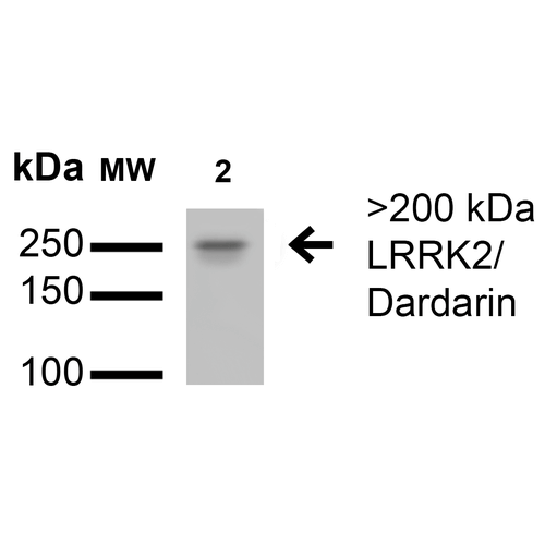 Western Blot analysis of Rat Brain Membrane showing detection of >200 kDa LRRK2/Dardarin protein using Mouse Anti-LRRK2/Dardarin Monoclonal Antibody, Clone S138-6 (56563). Lane 1: Molecular Weight Ladder. Lane 2: Rat Brain Membrane. Load: 15 µg. Block: 2% BSA and 2% Skim Milk in 1X TBST. Primary Antibody: Mouse Anti-LRRK2/Dardarin Monoclonal Antibody (56563) at 1:200 for 16 hours at 4°C. Secondary Antibody: Goat Anti-Mouse IgG: HRP at 1:1000 for 1 hour RT. Color Development: ECL solution for 6 min in RT. Predicted/Observed Size: >200 kDa.
 - -
- -
Antibody DetailsProduct DetailsReactive Species Human ⋅ Mouse ⋅ Rat Host Species Mouse Immunogen Fusion protein corresponding to aa 1-500 (N-terminus) of human LRRK2. This sequence is 83 Product Concentration 1.0 mg/ml Formulation PBS, pH 7.4, 0.1% sodium azide, 50% glycerol. State of Matter Liquid Product Preparation Purified by Protein G affinity chromatography Storage and Handling This product is stable for at least one (1) year at -20°C. Regulatory Status For in vitro investigational use only. Not intended for therapeutic or diagnostic procedures. Country of Origin USA Shipping Next Day 2-8°C Applications and Recommended Usage? Quality Tested by Leinco Immunoblotting: use at 1-5ug/mL. A band of >200kDa is detected.
Immunofluorescence: use at 10ug/mL. These are recommended concentrations. Endusers should determine optimal concentrations for their application. Each investigator should determine their own optimal working dilution for specific applications. See directions on lot specific datasheets, as information may periodically change. DescriptionDescriptionSpecificity This antibody recognizes human, mouse and rat LRRK2. Background LRRK2 is a large protein (>200kDa) with multiple domains including several ankyrin, leucine-rich, and WD40 repeats, a Raslike small GTPase family domain named Roc, and a kinase domain that is closely related to the RIP kinase domain. The LRRK2 gene is expressed in brain as well as in other tissues such as lung, liver and heart. LRRK2 might be central to the pathogenesis of several major neurodegenerative diseases associated with parkinsonism. LRRK2 is also known as Leucine-rich repeat serine/threonine-protein kinase 2, Dardarin, and PARK8. Function Serine/threonine-protein kinase which phosphorylates a broad range of proteins involved in multiple processes such as neuronal plasticity, autophagy, and vesicle trafficking (PubMed:20949042, PubMed:22012985, PubMed:26824392, PubMed:29125462, PubMed:28720718, PubMed:29127255, PubMed:30398148, PubMed:29212815, PubMed:30635421, PubMed:21850687, PubMed:23395371, PubMed:17114044, PubMed:24687852, PubMed:26014385, PubMed:25201882). Is a key regulator of RAB GTPases by regulating the GTP/GDP exchange and interaction partners of RABs through phosphorylation (PubMed:26824392, PubMed:28720718, PubMed:29127255, PubMed:30398148, PubMed:29212815, PubMed:29125462, PubMed:30635421). Phosphorylates RAB3A, RAB3B, RAB3C, RAB3D, RAB5A, RAB5B, RAB5C, RAB8A, RAB8B, RAB10, RAB12, RAB35, and RAB43 (PubMed:26824392, PubMed:28720718, PubMed:29127255, PubMed:30398148, PubMed:29212815, PubMed:29125462, PubMed:30635421, PubMed:23395371). Regulates the RAB3IP-catalyzed GDP/GTP exchange for RAB8A through the phosphorylation of 'Thr-72' on RAB8A (PubMed:26824392). Inhibits the interaction between RAB8A and GDI1 and/or GDI2 by phosphorylating 'Thr-72' on RAB8A (PubMed:26824392). Regulates primary ciliogenesis through phosphorylation of RAB8A and RAB10, which promotes SHH signaling in the brain (PubMed:29125462, PubMed:30398148). Together with RAB29, plays a role in the retrograde trafficking pathway for recycling proteins, such as mannose-6-phosphate receptor (M6PR), between lysosomes and the Golgi apparatus in a retromer-dependent manner (PubMed:23395371). Regulates neuronal process morphology in the intact central nervous system (CNS) (PubMed:17114044). Plays a role in synaptic vesicle trafficking (PubMed:24687852). Plays an important role in recruiting SEC16A to endoplasmic reticulum exit sites (ERES) and in regulating ER to Golgi vesicle-mediated transport and ERES organization (PubMed:25201882). Positively regulates autophagy through a calcium-dependent activation of the CaMKK/AMPK signaling pathway (PubMed:22012985). The process involves activation of nicotinic acid adenine dinucleotide phosphate (NAADP) receptors, increase in lysosomal pH, and calcium release from lysosomes (PubMed:22012985). Phosphorylates PRDX3 (PubMed:21850687). By phosphorylating APP on 'Thr-743', which promotes the production and the nuclear translocation of the APP intracellular domain (AICD), regulates dopaminergic neuron apoptosis (PubMed:28720718). Independent of its kinase activity, inhibits the proteosomal degradation of MAPT, thus promoting MAPT oligomerization and secretion (PubMed:26014385). In addition, has GTPase activity via its Roc domain which regulates LRRK2 kinase activity (PubMed:18230735, PubMed:26824392, PubMed:29125462, PubMed:28720718, PubMed:29212815). {PubMed:17114044, PubMed:18230735, PubMed:20949042, PubMed:21850687, PubMed:22012985, PubMed:23395371, PubMed:24687852, PubMed:25201882, PubMed:26014385, PubMed:26824392, PubMed:28720718, PubMed:29125462, PubMed:29127255, PubMed:29212815, PubMed:30398148, PubMed:30635421}. NCBI Gene Bank ID UniProt.org Research Area Neuroscience References & CitationsTechnical ProtocolsICC IF  Certificate of Analysis |
