

Anti-MMP 9 Antibody (29022)


Anti-MMP 9 Antibody (29022)
Product No.: 29022
- -
- -
Clone S51-82 Target MMP-9 Formats AvailableView All Product Type Monoclonal Alternate Names MMP-9, EC 3.4.24.35, 92 kDa gelatinase, 92 kDa type IV collagenase, Gelatinase B, GELB Isotype Mouse IgG2a Applications IHC , IP , WB , FCM , ICC/IF |
Data
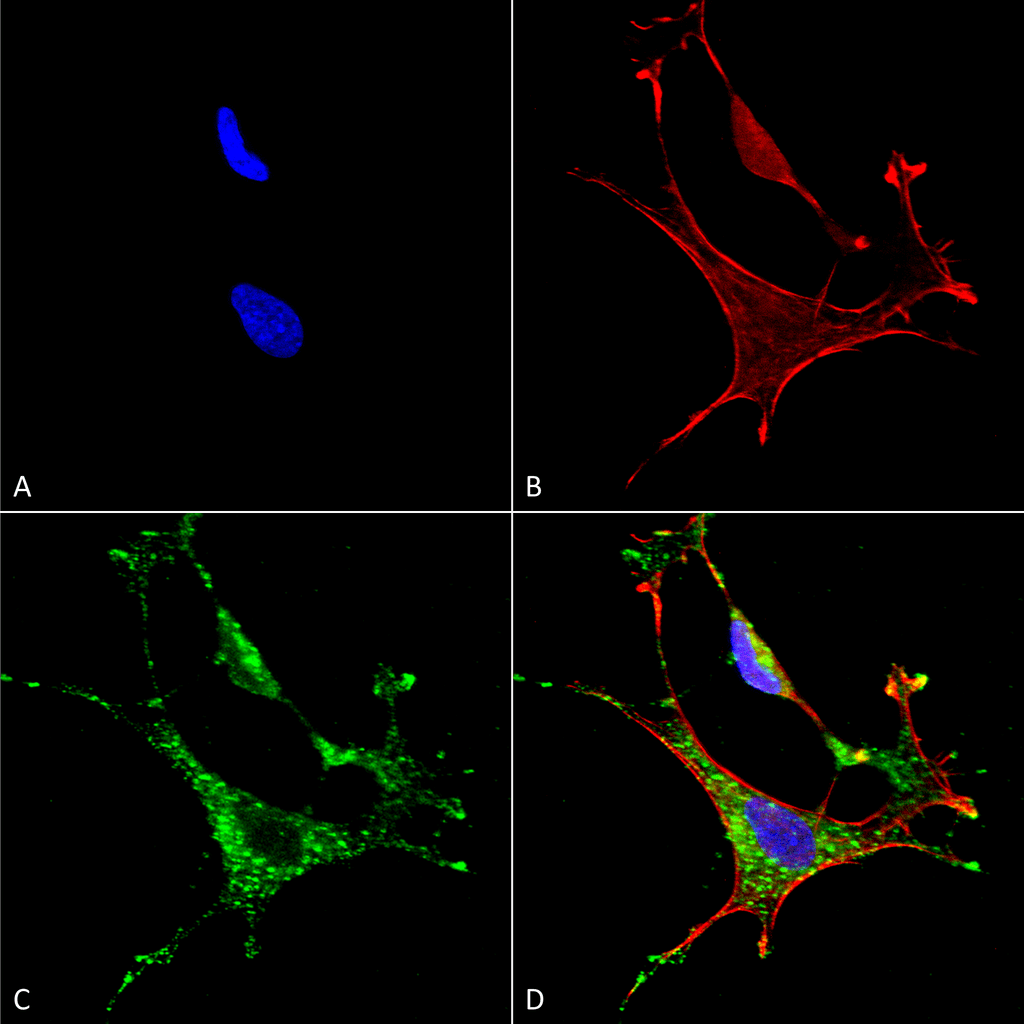 Immunocytochemistry/Immunofluorescence analysis using Mouse Anti-MMP9 Monoclonal Antibody, Clone S51-82 (29022). Tissue: Neuroblastoma cells (SH-SY5Y). Species: Human. Fixation: 4% PFA for 15 min. Primary Antibody: Mouse Anti-MMP9 Monoclonal Antibody (29022) at 1:50 for overnight at 4°C with slow rocking. Secondary Antibody: AlexaFluor 488 at 1:1000 for 1 hour at RT. Counterstain: Phalloidin-iFluor 647 (red) F-Actin stain; Hoechst (blue) nuclear stain at 1:800, 1.6mM for 20 min at RT. (A) Hoechst (blue) nuclear stain. (B) Phalloidin-iFluor 647 (red) F-Actin stain. (C) MMP9 Antibody (D) Composite.
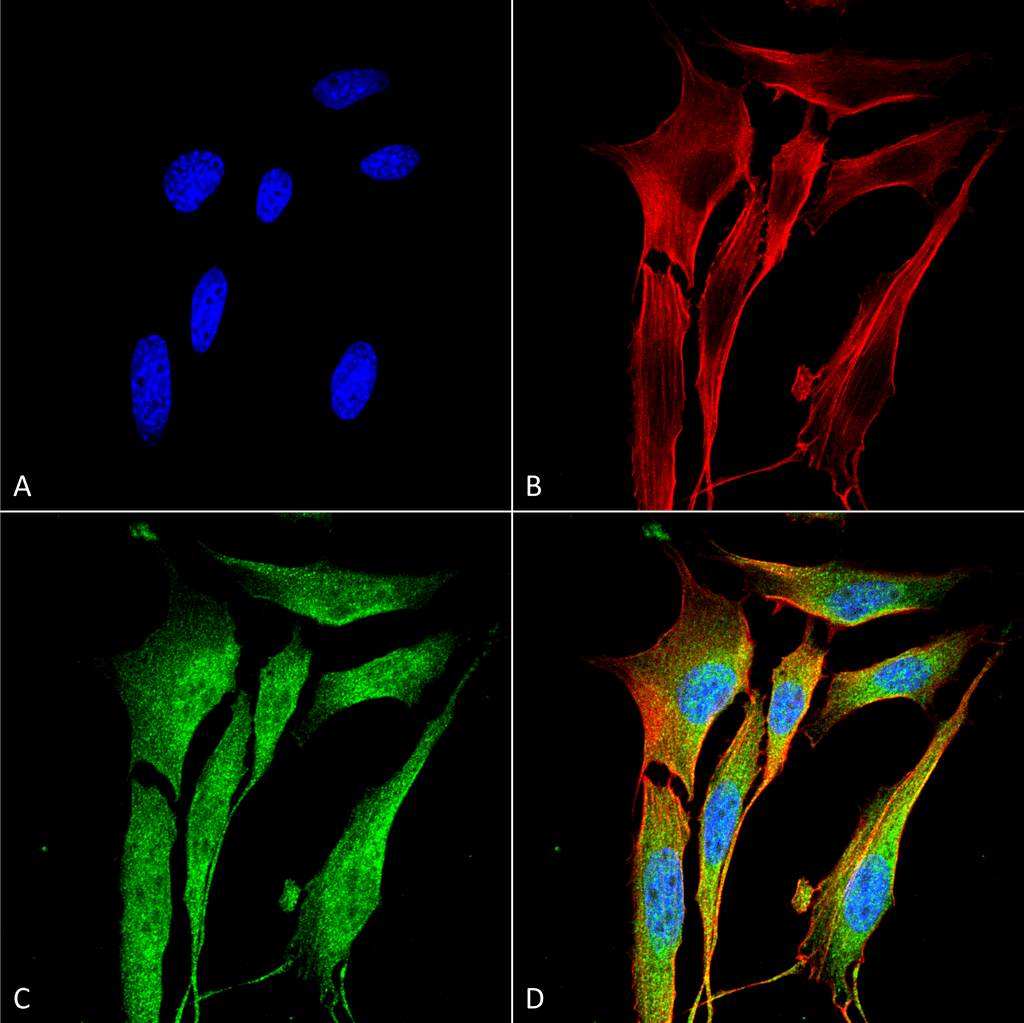 Immunocytochemistry/Immunofluorescence analysis using Mouse Anti-MMP9 Monoclonal Antibody, Clone S51-82 (29022). Tissue: NIH 3T3 (NIH 3T3). Species: Mouse. Fixation: 4% Formaldehyde for 15 min at RT. Primary Antibody: Mouse Anti-MMP9 Monoclonal Antibody (29022) at 1:100 for 60 min at RT. Secondary Antibody: Goat Anti-Mouse ATTO 488 at 1:200 for 60 min at RT. Counterstain: Phalloidin Texas Red F-Actin stain; DAPI (blue) nuclear stain at 1:1000, 1:5000 for 60 min at RT, 5 min at RT. Localization: Cytoplasm . Magnification: 60X. (A) DAPI (blue) nuclear stain. (B) Phalloidin Texas Red F-Actin stain. (C) MMP9 Antibody. (D) Composite.
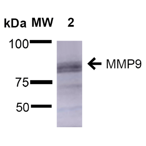 Western Blot analysis of Rat Brain showing detection of ~92 kDa and ~82 kDa (pro and active) MMP9 protein using Mouse Anti-MMP9 Monoclonal Antibody, Clone S51-82 (29022). Lane 1: Molecular Weight Ladder (MW). Lane 2: Rat Brain. Load: 15 µg. Block: 5% Skim Milk in 1X TBST. Primary Antibody: Mouse Anti-MMP9 Monoclonal Antibody (29022) at 1:1000 for 2 hours at RT. Secondary Antibody: Goat Anti-Mouse IgG: HRP at 1:2000 for 60 min at RT. Color Development: ECL solution for 5 min at RT. Predicted/Observed Size: ~92 kDa and ~82 kDa (pro and active).
 - -
- -
Antibody DetailsProduct DetailsReactive Species Human ⋅ Mouse ⋅ Rat Host Species Mouse Immunogen Fusion protein corresponding to aa 1-708 (full-length) of rat MMP 9 (accession no. NP_112317). Product Concentration 1.0 mg/ml Formulation PBS, pH 7.4, 50% glycerol, 0.09% sodium azide.Purified by Protein G affinity chromatography. State of Matter Liquid Product Preparation Purified by Protein G affinity chromatography Storage and Handling This product is stable for at least 1 year at -20°C. Freeze in multiple aliquots to avoid repeated freeze-thaw cycles. Regulatory Status For in vitro investigational use only. Not for
use in therapeutic or diagnostic procedures. Country of Origin USA Shipping Next Day 2-8°C Applications and Recommended Usage? Quality Tested by Leinco Immunoblotting: use at 1-2ug/mL. Bands of ~92 and 82kDa (proenzyme and active enzyme) are detected.
Immunohistochemistry: use at 1-5ug/mL. These are recommended concentrations. Enduser should determine optimal concentrations for their applications. Positive control: rat brain lysate. Each investigator should determine their own optimal working dilution for specific applications. See directions on lot specific datasheets, as information may periodically change. DescriptionDescriptionSpecificity This antibody recognizes human, mouse, and rat MMP 9. Background MMP 9 is involved in the breakdown of extracellular matrix in embryonic development, reproduction, wound healing, and tissue remodeling as well as in disease processes such as arthiritis and metastasis. Once synthesized, MMPs exist as latent proenzymes. Maximum MMP activity requires proteolytic cleavage of the proenzyme to generate active MMP by releasing the inhibitory propeptide domain from the full-length protein. Function Matrix metalloproteinase that plays an essential role in local proteolysis of the extracellular matrix and in leukocyte migration (By similarity). Could play a role in bone osteoclastic resorption (By similarity). Cleaves KiSS1 at a Gly-|-Leu bond (By similarity). Cleaves NINJ1 to generate the Secreted ninjurin-1 form (By similarity). Cleaves type IV and type V collagen into large C-terminal three quarter fragments and shorter N-terminal one quarter fragments. Degrades fibronectin but not laminin or Pz-peptide (By similarity). {UniProtKB:P14780, UniProtKB:P41245}. NCBI Gene Bank ID UniProt.org Research Area Matrix/Metalloproteins References & CitationsTechnical Protocols   FCM ICC/IF Certificate of Analysis |
Formats Available
 Products are for research use only. Not for use in diagnostic or therapeutic procedures.