

Anti-Mouse Alpha Synuclein Mouse Monoclonal Antibody (56625)

Anti-Mouse Alpha Synuclein Mouse Monoclonal Antibody (56625)
Product No.: 56625
- -
- -
Clone 10H7 Target Alpha Synuclein Formats AvailableView All Product Type Monoclonal Alternate Names Alpha Synuclein, Non-A beta component of AD amyloid, Non-A4 component of amyloid precursor, NACP, SNCA, PARK1, PARK 1, alphaSYN, PARK 4, PARK4, Parkinson disease familial 1, Parkinson disease (autosomal dominant, Lewy body) 4, SYN Isotype Mouse IgG1 Applications ELISA , ICC , IF , WB |
Data
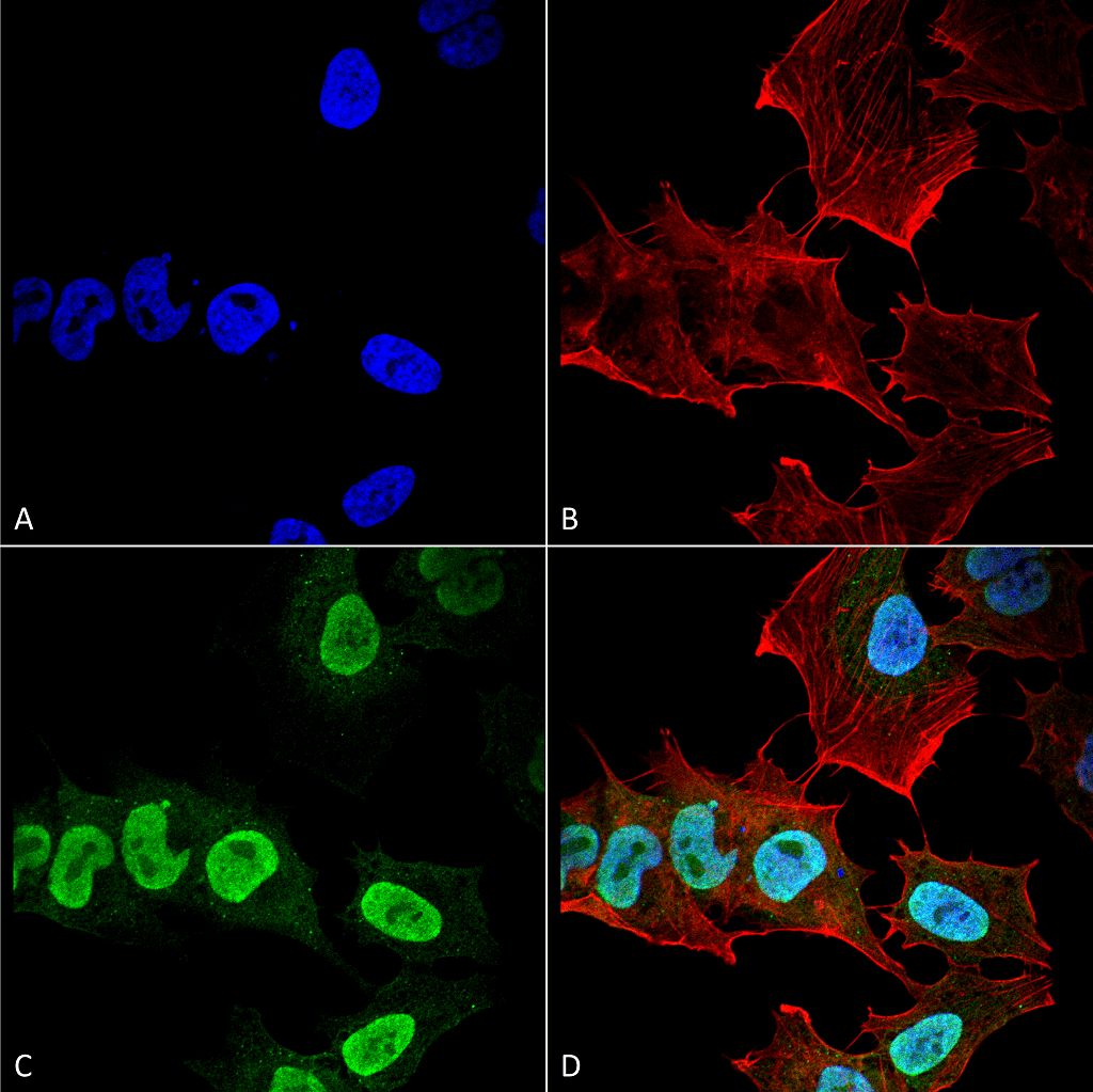 Immunocytochemistry/Immunofluorescence analysis using Mouse Anti-Alpha Synuclein Monoclonal Antibody, Clone 10H7 . Tissue: Neuroblastoma cell line (SK-N-BE). Species: Human. Fixation: 4% Formaldehyde for 15 min at RT. Primary Antibody: Mouse Anti-Alpha Synuclein Monoclonal Antibody at 1:100 for 60 min at RT. Secondary Antibody: Goat Anti-Mouse ATTO 488 at 1:200 for 60 min at RT. Counterstain: Phalloidin Texas Red F-Actin stain; DAPI (blue) nuclear stain at 1:1000, 1:5000 for 60 min at RT, 5 min at RT. Localization: Cytoplasm: weak; Nucleus: Med. Magnification: 60X. (A) DAPI (blue) nuclear stain. (B) Phalloidin Texas Red F-Actin stain. (C) Alpha Synuclein Antibody. (D) Composite.
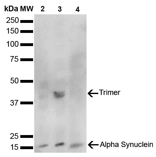 Western Blot analysis of Human, Mouse, Rat Brain showing detection of 14 kDa Alpha Synuclein protein using Mouse Anti-Alpha Synuclein Monoclonal Antibody, Clone 10H7 . Lane 1: Molecular Weight Ladder (MW). Lane 2: Mouse Brain cell lysate. Lane 3: Rat brain cell lysate. Lane 4: Human brain cell lysate. Load: 15 µg. Block: 5% Skim Milk in 1X TBST. Primary Antibody: Mouse Anti-Alpha Synuclein Monoclonal Antibody at 1:1000 for 2 hours at RT. Secondary Antibody: Goat Anti-Mouse HRP:IgG at 1:3000 for 1 hour at RT. Color Development: ECL solution (Super Signal West Pico) for 5 min in RT. Predicted/Observed Size: 14 kDa. Other Band(s): ~40 kDa (trimer).
 - -
- -
Antibody DetailsProduct DetailsReactive Species Human ⋅ Mouse ⋅ Rat Host Species Mouse Immunogen Mouse alpha synuclein aggregate Product Concentration 1 mg/mL Formulation PBS pH 7.4, 50% glycerol, 0.09% Sodium azide State of Matter Liquid Product Preparation Protein G Purified Storage and Handling This antibody is stable for at least one (1) year at -20°C. Avoid multiple freeze-thaw cycles. Regulatory Status Research Use Only Country of Origin USA Shipping Next Day 2-8°C Applications and Recommended Usage? Quality Tested by Leinco WB (1:1000); DB (1:1000); ICC/IF (1:100); ELISA (1:1000); optimal dilutions for assays should be determined by the user. Each investigator should determine their own optimal working dilution for specific applications. See directions on lot specific datasheets, as information may periodically change. DescriptionDescriptionSpecificity Binds within the 101-130 aa region of human alpha synuclein, determined using Dot blot and ELISA with corresponding peptides. Detects ~14 kDa band corresponding to Alpha synuclein. Band at ~40 kDa is a trimer. Does not detect truncated alpha Synuclein (aa 1-114). Background Alpha-Synuclein (SNCA) is expressed predominantly in the brain, where it is concentrated in presynaptic nerve terminals (1). Alpha-synuclein is highly expressed in the mitochondria of the olfactory bulb, hippocampus, striatum and thalamus (2). Functionally, it has been shown to significantly interact with tubulin (3), and may serve as a potential microtubule-associated protein. It has also been found to be essential for normal development of the cognitive functions; inactivation may lead to impaired spatial learning and working memory (4). SNCA fibrillar aggregates represent the major non A-beta component of Alzheimers disease amyloid plaque, and a major component of Lewy body inclusions, and Parkinson's disease. Parkinson's disease (PD) is a common neurodegenerative disorder characterized by the progressive accumulation in selected neurons of protein inclusions containing alpha-synuclein and ubiquitin (5, 6). NCBI Gene Bank ID UniProt.org Research Area Neuroscience . Alzheimer's Disease . Neurodegeneration . Parkinson's Disease . Synuclein . Tangles & Tau References & CitationsTechnical Protocols ICC IF  Certificate of Analysis |
