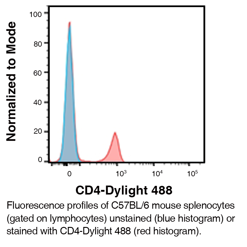
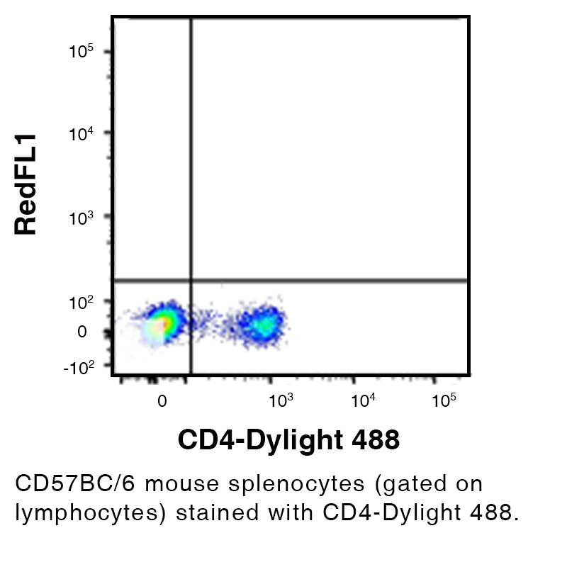
Anti-Mouse CD4 (Clone GK1.5) – DyLight® 488
Anti-Mouse CD4 (Clone GK1.5) – DyLight® 488
Product No.: C1636
- -
- -
Clone GK1.5 Target CD4 Formats AvailableView All Product Type Monoclonal Antibody Alternate Names CD4mut, L3T4, Ly-4, Cd4, CD4 Antigen Isotype IgG2b Applications FC , ICC , IHC FF |
Data
- -
- -
Antibody DetailsProduct DetailsReactive Species Mouse Host Species Rat Product Concentration 0.2 mg/ml Formulation This DyLight® 488 conjugate is formulated in 0.01 M phosphate buffered saline (150 mM NaCl) PBS pH 7.4, 1% BSA and 0.09% sodium azide as a preservative. Storage and Handling This DyLight® 488 conjugate is stable when stored at 2-8°C. Do not freeze. Country of Origin USA Shipping Next Day 2-8°C Excitation Laser Blue Laser (493 nm) RRIDAB_2828691 Applications and Recommended Usage? Quality Tested by Leinco FC The suggested concentration for this GK1.5 antibody for staining cells in flow cytometry is ≤ 0.5 μg per 106 cells in a volume of 100 μl. Titration of the reagent is recommended for optimal performance for each application. Additional Applications Reported In Literature ? IHC-FF ICC Each investigator should determine their own optimal working dilution for specific applications. See directions on lot specific datasheets, as information may periodically change. DescriptionDescriptionSpecificity Rat Anti-Mouse CD4 (Clone GK1.5) recognizes an epitope on Mouse CD4. This monoclonal antibody was purified using multi-step affinity chromatography methods such as Protein A or G depending on the species and isotype. Background CD4 (cluster of differentiation 4) is a glycoprotein expressed on the surface of T helper cells, regulatory T cells, monocytes, macrophages, and dendritic cells. CD4 interacts with class II molecules of the major histocompatibility complex (MHC) enhancing the signal for T-cell activation.1 Antigen Distribution The L3T4 antigen is expressed by the helper/inducer subset of mouse T-cells. The antigen is present on approximately 80% of thymocytes, 20% of spleen cells and 60% of lymph node cells. The expression of L3T4 correlates with class II MHC antigen reactivity on cloned T-cell lines. Ligand/Receptor MHC class II molecule PubMed NCBI Gene Bank ID UniProt.org Research Area Immunology References & Citations1. Hendrickson WA et al. (1994) Structure 2: 59 Technical ProtocolsCertificate of Analysis |
Related Products
- -
- -
Formats Available
- -
- -
Prod No. | Description |
|---|---|
C1840 | |
C211 | |
C214 | |
C212 | |
C213 | |
C338 | |
C1640 | |
C1636 | |
C1333 | |
C1638 | |
C1637 | |
C2838 |


