

Anti-Mouse E-Cadherin

Data
- -
- -
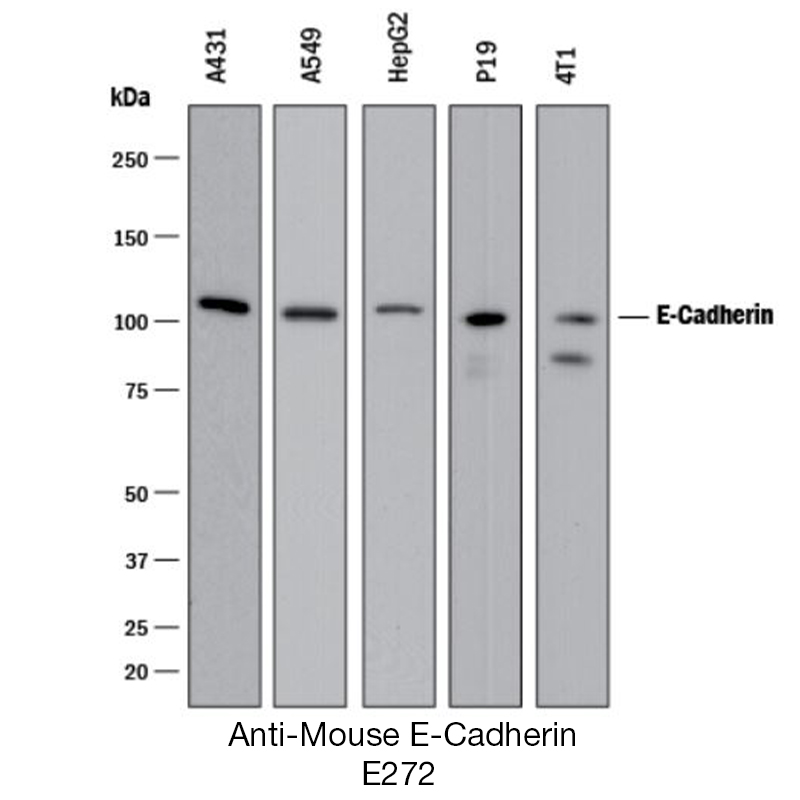
Antibody DetailsProduct DetailsReactive Species Mouse Host Species Goat Immunogen NS0-Derived Recombinant Mouse E-Cadherin Extracellular Domain Endotoxin Level <0.1 EU/µg as determined by the LAL method Formulation This antigen affinity purified polyclonal antibody has been 0.2 µm filtered and lyophilized from modified Dulbecco’s phosphate buffered saline (1X PBS) pH 7.2 – 7.3 containing 5.0% w/v trehalose with no calcium, magnesium, or preservatives present. State of Matter Lyophilized Storage and Handling The lyophilized antigen affinity purified polyclonal antibody can be stored desiccated at -20°C to -70°C for twelve months from date of receipt. The reconstituted antibody can be stored for at least four weeks at 2-8°C. For long-term storage of the reconstituted antibody, aseptically aliquot into working volumes and store at -20°C to -70°C in a manual defrost freezer. Avoid Repeated Freeze Thaw Cycles. No detectable loss of activity was observed after six months. Country of Origin USA Shipping Next Day Ambient RRIDAB_2830044 Applications and Recommended Usage? Quality Tested by Leinco Flow Cytometry: It is recommended to use the indirect method for signal enhancement when enumerating cells expressing E-Cadherin. A suggested method would be to stain cells expressing E-Cadherin with 0.50 µg per 1 - 2.5 x 10⁶ cells in a total staining volume of ≤200 µl followed by PN:G641. Western Blotting: To detect Mouse E-Cadherin this polyclonal antibody can be used at a concentration of 0.5 µg/ml. This polyclonal antibody should be used in conjunction with compatible second-step reagents such as PN:G505 and a chromogenic substrate such as PN:T343. The detection limit for Mouse E-Cadherin is 1 ng/lane under either reducing or non-reducing conditions. The sensitivity of detection may increase up to 50 fold when a chemiluminescent substrate is used. Additional Applications Reported In Literature ? IHC (NBF/Par.): This antibody should give satisfactory staining results when used at a concentration of 1.7 - 5 µg/ml. The recommended secondary antibody for IHC is PN:G505. For chromogenic detection with high signal and low background use PN:D100 or PN:K107. Immunocytochemistry: Suitable for use at concentration of 5-15 µg/mL. CyTOF-ready: Ready to be labeled using established conjugation methods. No BSA or other carrier proteins that could interfere with conjugation. Each investigator should determine their own optimal working dilution for specific applications. See directions on lot specific datasheets, as information may periodically change. DescriptionDescriptionSpecificity Goat Anti-Mouse E-Cadherin (E-CAD) recognizes Mouse E-CAD. This antigen affinity purified polyclonal antibody was purified using a proprietary chromatographic technique that includes covalently immobilizing the antigen proteins or peptides to agarose based beads. This purification method enhances specificity, reduces nonspecific binding of extraneous IgG and provides you with the most reliable reagent available for your early discovery research. Background Cadherin 1, type 1, E-cadherin (epithelial), also known as CDH1 and CD32, is a tumor suppressor gene. 1, 2 The encoded protein is a calcium dependent cell-cell adhesion glycoprotein comprised of five extracellular cadherin repeats, a transmembrane region and a highly conserved cytoplasmic tail. E-cadherin-mediated cell-cell adhesion pathway may represent a novel chemotherapeutic target for bladder cancer, prostate cancer, and renal-cell carcinoma. 3 PubMed NCBI Gene Bank ID UniProt.org References & Citations1. Christofori, G. et al.(1998) Am. J. Hum. Genet. 63: 1588 2. Gumbiner, BM. et al.(2003) J. Cell Biol.161: 1191 3. Isaacs, WB. et al.(1995) World J Urol.13:364 Technical ProtocolsCyTOF®  ICC IHC FFPE  Certificate of Analysis |
