

Anti-Neuroligin-3 Antibody (56552)

Anti-Neuroligin-3 Antibody (56552)
Product No.: 56552
- -
- -
Clone S110-29 Target Neuroligin-3 Formats AvailableView All Product Type Monoclonal Alternate Names Gliotactin homolog Isotype Mouse IgG1 Applications ICC , IF , IHC , WB |
Data
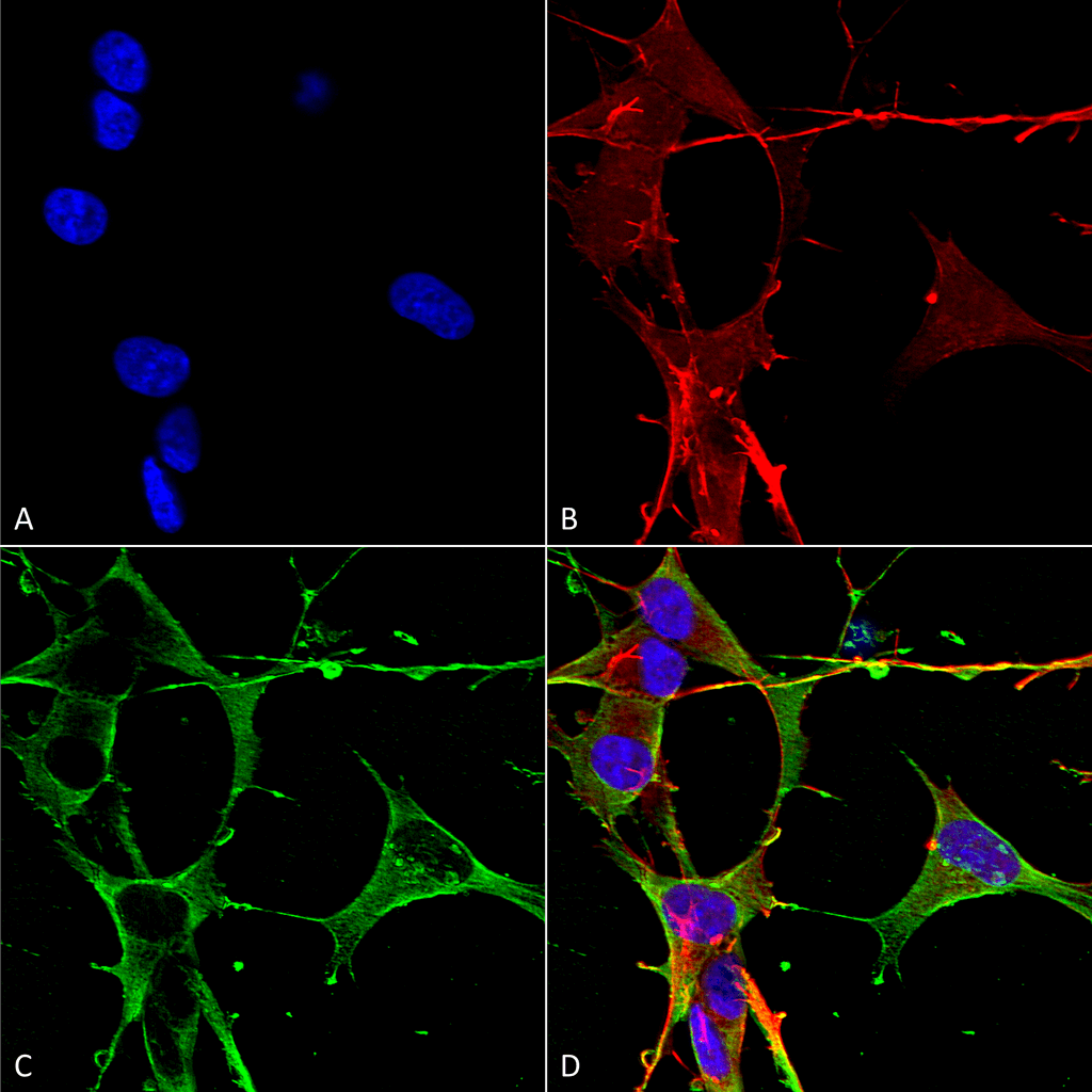 Immunocytochemistry/Immunofluorescence analysis using Mouse Anti-Neuroligin 3 Monoclonal Antibody, Clone S110-29 (56552). Tissue: Neuroblastoma cells (SH-SY5Y). Species: Human. Fixation: 4% PFA for 15 min. Primary Antibody: Mouse Anti-Neuroligin 3 Monoclonal Antibody (56552) at 1:50 for overnight at 4°C with slow rocking. Secondary Antibody: AlexaFluor 488 at 1:1000 for 1 hour at RT. Counterstain: Phalloidin-iFluor 647 (red) F-Actin stain; Hoechst (blue) nuclear stain at 1:800, 1.6mM for 20 min at RT. (A) Hoechst (blue) nuclear stain. (B) Phalloidin-iFluor 647 (red) F-Actin stain. (C) Neuroligin 3 Antibody (D) Composite.
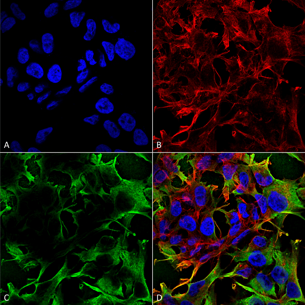 Immunocytochemistry/Immunofluorescence analysis using Mouse Anti-Neuroligin 3 Monoclonal Antibody, Clone S110-29 (56552). Tissue: Neuroblastoma cell line (SK-N-BE). Species: Human. Fixation: 4% Formaldehyde for 15 min at RT. Primary Antibody: Mouse Anti-Neuroligin 3 Monoclonal Antibody (56552) at 1:100 for 60 min at RT. Secondary Antibody: Goat Anti-Mouse ATTO 488 at 1:100 for 60 min at RT. Counterstain: Phalloidin Texas Red F-Actin stain; DAPI (blue) nuclear stain at 1:1000, 1:5000 for 60min RT, 5min RT. Localization: Cell Membrane, Cell Junction, Synapse . Magnification: 60X. (A) DAPI (blue) nuclear stain. (B) Phalloidin Texas Red F-Actin stain. (C) Neuroligin 3 Antibody. (D) Composite.
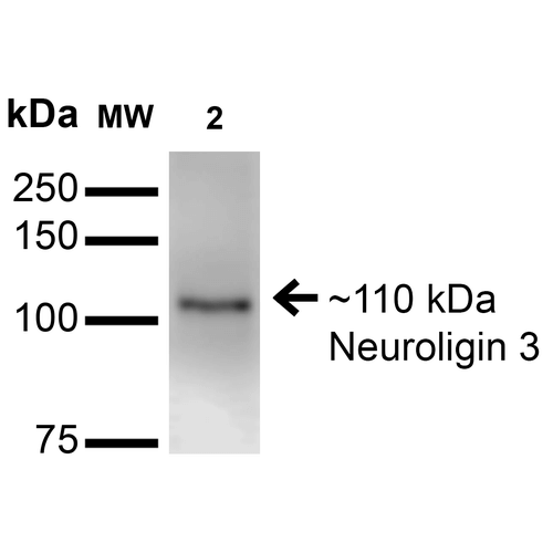 Western Blot analysis of Mouse Brain Membrane showing detection of ~110 kDa Neuroligin 3 protein using Mouse Anti-Neuroligin 3 Monoclonal Antibody, Clone S110-29 (56552). Lane 1: Molecular Weight Ladder. Lane 2: Mouse Brain Membrane. Load: 15 µg. Block: 2% BSA and 2% Skim Milk in 1X TBST. Primary Antibody: Mouse Anti-Neuroligin 3 Monoclonal Antibody (56552) at 1:200 for 16 hours at 4°C. Secondary Antibody: Goat Anti-Mouse IgG: HRP at 1:1000 for 1 hour RT. Color Development: ECL solution for 6 min in RT. Predicted/Observed Size: ~110 kDa.
 - -
- -
Antibody DetailsProduct DetailsReactive Species Human ⋅ Mouse ⋅ Rat Host Species Mouse Immunogen Fusion protein corresponding to aa 730-848 (intracellular C-terminus) of rat Neuroligin- 3. This sequence is 99 Product Concentration 1.0 mg/ml Formulation PBS, pH 7.4, 0.1% sodium azide, 50% glycerol. State of Matter Liquid Product Preparation Purified by Protein G affinity chromatography Storage and Handling This product is stable for at least one (1) year at -20°C. Regulatory Status For in vitro investigational use only. Not intended for therapeutic or diagnostic procedures. Country of Origin USA Shipping Next Day 2-8°C Applications and Recommended Usage? Quality Tested by Leinco Immunoblotting: use at 1-5ug/mL. A band of ~110kDa is detected.
Immunofluorescence: use at 10ug/mL. These are recommended concentrations. Endusers should determine optimal concentrations for their application. Each investigator should determine their own optimal working dilution for specific applications. See directions on lot specific datasheets, as information may periodically change. DescriptionDescriptionSpecificity This antibody recognizes human, mouse and rat Neuroligin-3. It does not cross-react with Neuroligin-1, -2, or -4. Background Neuroligin-3 is a neuronal cell surface protein involved in cell-cell-interactions via its interactions with neurexin family members. It plays a role in synapse function and synaptic signal transmission, and may mediate its effects by clustering other synaptic proteins. It may also promote the initial formation of synapses and play a role in glia-glia or glia-neuron interactions in the developing peripheral nervous system. Mutations in this gene may be associated with autism and Asperger syndrome. Multiple transcript variants encoding distinct isoforms have been identified for this gene. Function Cell surface protein involved in cell-cell-interactions via its interactions with neurexin family members. Plays a role in synapse function and synaptic signal transmission, and probably mediates its effects by recruiting and clustering other synaptic proteins. May promote the initial formation of synapses, but is not essential for this. May also play a role in glia-glia or glia-neuron interactions in the developing peripheral nervous system. {PubMed:17897391}. NCBI Gene Bank ID UniProt.org Research Area Neuroscience References & CitationsTechnical ProtocolsICC IF   Certificate of Analysis |
