

Anti-p53 Antibody (3347)

Data
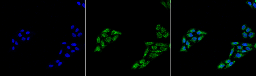 Immunocytochemistry/Immunofluorescence analysis using Rabbit Anti-p53 Polyclonal Antibody (3347). Tissue: Cervical cancer cell line (HeLa). Species: Human. Fixation: 4% Formaldehyde for 15 min at RT. Primary Antibody: Rabbit Anti-p53 Polyclonal Antibody (3347) at 1:100 for 60 min at RT. Secondary Antibody: Goat Anti-Rabbit ATTO 488 at 1:100 for 60 min at RT. Counterstain: DAPI (blue) nuclear stain at 1:5000 for 5 min RT. Localization: Cytoplasm, PML body, Endoplasmic Reticulum. Magnification: 40X. (A) DAPI (blue) nuclear stain (B) Phalloidin Texas Red F-Actin stain (C) p53 Antibody (D) Composite.
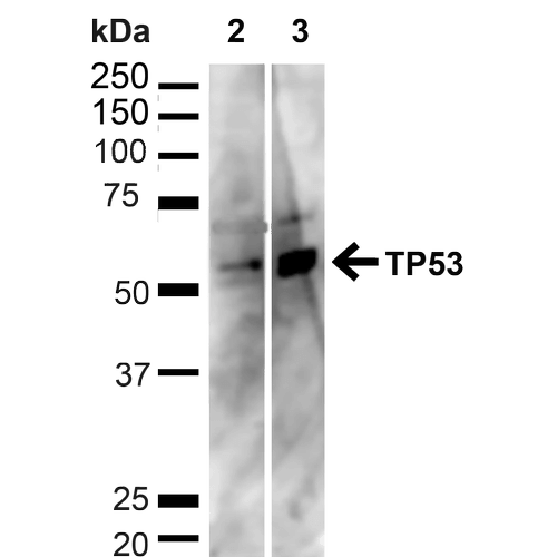 Western blot analysis of Human HeLa and HEK293T cell lysates showing detection of ~43.7kDa p53 protein using Rabbit Anti-p53 Polyclonal Antibody (3347). Lane 1: MW Ladder. Lane 2: Human HeLa (20 µg). Lane 3: Human 293T (20 µg). Load: 20 µg. Block: 5% milk + TBST for 1 hour at RT. Primary Antibody: Rabbit Anti-p53 Polyclonal Antibody (3347) at 1:1000 for 1 hour at RT. Secondary Antibody: Goat Anti-Rabbit: HRP at 1:2000 for 1 hour at RT. Color Development: TMB solution for 12 min at RT. Predicted/Observed Size: ~43.7kDa.
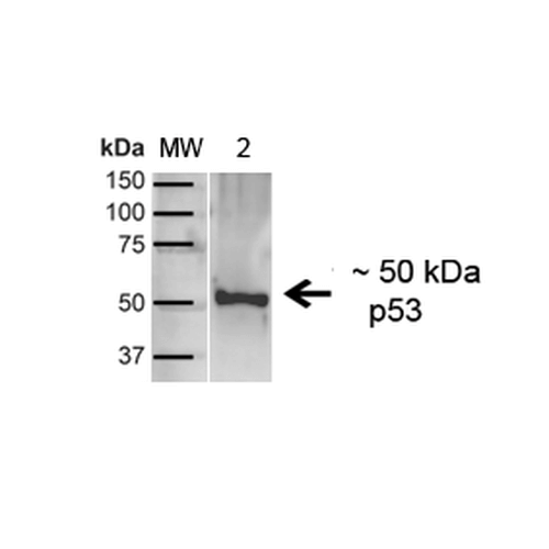 Western blot analysis of Human A431 showing detection of ~43.7kDa p53 protein using Rabbit Anti-p53 Polyclonal Antibody (3347). Lane 1: MW Ladder. Lane 2: Human A431 (20 µg). Load: 20 µg. Block: 5% milk + TBST for 1 hour at RT. Primary Antibody: Rabbit Anti-p53 Polyclonal Antibody (3347) at 1:1000 for 1 hour at RT. Secondary Antibody: Goat Anti-Rabbit: HRP at 1:2000 for 1 hour at RT. Color Development: TMB solution for 12 min at RT. Predicted/Observed Size: ~43.7kDa.
 - -
- -
Antibody DetailsProduct DetailsReactive Species Human Host Species Rabbit Immunogen Synthetic peptide corresponding to amino acids at the C-terminus of human p53. Product Concentration Lot Specific Formulation PBS, pH 7.4, 50% glycerol, 0.09% sodium azide. State of Matter Liquid Product Preparation Purified by Protein A affinity chromatography Storage and Handling This antibody is stable for at least one (1) year at -20°C. Regulatory Status For in vitro investigational use only. Not intended for therapeutic or diagnostic applications. Country of Origin USA Shipping Next Day 2-8°C Applications and Recommended Usage? Quality Tested by Leinco Immunoblotting: use at 1ug/mL. A band of ~53kDa is detected.
Immunofluorescence: use at 10ug/mL. These are recommended concentrations. Endusers should determine optimal concentrations for their applications. Each investigator should determine their own optimal working dilution for specific applications. See directions on lot specific datasheets, as information may periodically change. DescriptionDescriptionSpecificity This antibody recognizes human p53. Background Tumor protein p53, also known as p53, functions as a tumor suppressor, and, as such, p53 has been described as “the guardian of the genome†because of its role in preventing genome mutations. The name p53 describes the apparent molecular mass of the protein as observed in SDS-PAGE analysis. The actual mass of full-length p53, based on the sum of masses of the amino acid residues, is 43.7kDa. The difference is due to the number of proline residues in the protein which slow its migration on SDS-PAGE, thus making it appear heavier than it actually is. The TP53 gene is the most frequently mutated gene (>50%) in human cancer. Function Acts as a tumor suppressor in many tumor types; induces growth arrest or apoptosis depending on the physiological circumstances and cell type. Involved in cell cycle regulation as a trans-activator that acts to negatively regulate cell division by controlling a set of genes required for this process. One of the activated genes is an inhibitor of cyclin-dependent kinases. Apoptosis induction seems to be mediated either by stimulation of BAX and FAS antigen expression, or by repression of Bcl-2 expression. Its pro-apoptotic activity is activated via its interaction with PPP1R13B/ASPP1 or TP53BP2/ASPP2 (PubMed:12524540). However, this activity is inhibited when the interaction with PPP1R13B/ASPP1 or TP53BP2/ASPP2 is displaced by PPP1R13L/iASPP (PubMed:12524540). In cooperation with mitochondrial PPIF is involved in activating oxidative stress-induced necrosis; the function is largely independent of transcription. Induces the transcription of long intergenic non-coding RNA p21 (lincRNA-p21) and lincRNA-Mkln1. LincRNA-p21 participates in TP53-dependent transcriptional repression leading to apoptosis and seems to have an effect on cell-cycle regulation. Implicated in Notch signaling cross-over. Prevents CDK7 kinase activity when associated to CAK complex in response to DNA damage, thus stopping cell cycle progression. Isoform 2 enhances the transactivation activity of isoform 1 from some but not all TP53-inducible promoters. Isoform 4 suppresses transactivation activity and impairs growth suppression mediated by isoform 1. Isoform 7 inhibits isoform 1-mediated apoptosis. Regulates the circadian clock by repressing CLOCK-ARNTL/BMAL1-mediated transcriptional activation of PER2 (PubMed:24051492). {PubMed:11025664, PubMed:12524540, PubMed:12810724, PubMed:15186775, PubMed:15340061, PubMed:17317671, PubMed:17349958, PubMed:19556538, PubMed:20673990, PubMed:20959462, PubMed:22726440, PubMed:24051492, PubMed:9840937}. NCBI Gene Bank ID UniProt.org Research Area Cancer Research References & CitationsTechnical ProtocolsICC IF  Certificate of Analysis |
