

Anti-Rhodopsin (Clone 4D2) Antibody

Anti-Rhodopsin (Clone 4D2) Antibody
Product No.: 56481
- -
- -
Clone 4D2 Target Rhodopsin Formats AvailableView All Product Type Monoclonal Isotype Mouse IgG1 Applications ELISA , ICC , IF , IHC , IP , WB |
Data
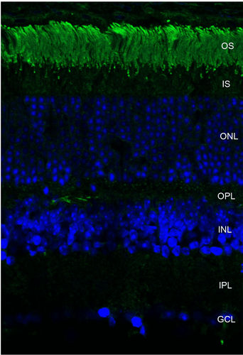 Immunohistochemistry analysis using Mouse Anti-Rhodopsin Monoclonal Antibody, Clone 4D2 (56481). Tissue: retina. Species: Mouse. Primary Antibody: Mouse Anti-Rhodopsin Monoclonal Antibody (56481) at 1:1000. Secondary Antibody: FITC Goat Anti-Mouse (green). Counterstain: DAPI (blue) nuclear stain. Localization: Staining of photoreceptor outer segment (OS). Other layers of the retina: IS – inner segment; ONL – outer nuclear layer; OPL – outer plexiform layer; INL – inner nuclear layer; IPL – inner plexiform layer; GCL – ganglion cell layer.
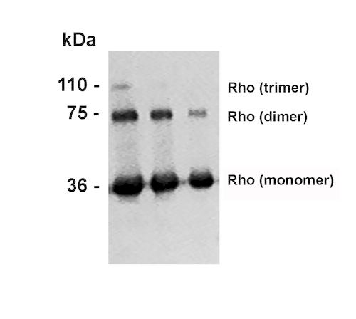 Western Blot analysis of Bovine photoreceptor membranes showing detection of Rhodopsin protein using Mouse Anti-Rhodopsin Monoclonal Antibody, Clone 4D2 (56481). Lane 1: MW ladder. Lane 2: 10 ug. Lane 3: 5 ug. Lane 4: 2.5 ug. Primary Antibody: Mouse Anti-Rhodopsin Monoclonal Antibody (56481) at 1:1000.
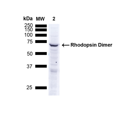 Western Blot analysis of Human A549 cells showing detection of ~38.9kDa Rhodopsin protein using Mouse Anti-Rhodopsin Monoclonal Antibody, Clone 4D2 (56481). Lane 1: MW ladder. Lane 2: Human A549 Cells 15 ug). Load: 15 ug. Block: 5% Skim Milk Powder in TBST. Primary Antibody: Mouse Anti-Rhodopsin Monoclonal Antibody (56481) at 1:1000 for 2.5 hours at RT with shaking . Secondary Antibody: Goat anti-mouse IgG:HRP at 1:1000 for 1 hour at RT with shaking . Color Development: Chemiluminescent for HRP (Moss) for 5 min in RT. Predicted/Observed Size: ~38.9kDa. Other Band(s): Band appears at ~75 kDa indicating detection of the Rhodopsin dimer.
 - -
- -
Antibody DetailsProduct DetailsReactive Species Amphibian ⋅ Avian ⋅ Fish ⋅ Mammals Host Species Mouse Immunogen Bovine rhodopsin Product Concentration Lot Specific Formulation PBS, pH 7.4; 50% glycerol, 0.09% sodium azide. State of Matter Liquid Product Preparation Purified by Protein G affinity chromatography Storage and Handling This antibody is stable for at least one (1) year at -20°C. Avoid repeated freezing and thawing. Regulatory Status For in vitro investigational use only. Not for use in therapeutic or diagnostic procedures. Country of Origin USA Shipping Next Day 2-8°C Applications and Recommended Usage? Quality Tested by Leinco Immunoblotting: use at 1-10ug/mL. A band of ~40kDa is detected.
Immunohistochemistry: use at 1-10ug/mL, paraffin-embedded and frozen. Immunoprecipitation: use at 1-2ug per 100-500ug of protein. These are recommended concentrations. User should determine optimal concentrations for their application. Positive control: Rat eye lysate Each investigator should determine their own optimal working dilution for specific applications. See directions on lot specific datasheets, as information may periodically change. DescriptionDescriptionSpecificity This antibody recognizes the N-terminus of Rhodopsin from all mammalian species tested (human, mouse, rat, bovine, porcine), and most fish, birds, and
amphibians. Background Rhodopsin is comprised of the protein opsin and a reversibly covalently bound cofactor, retinal. Opsin, made up of seven membrane-embedded alpha helices, binds retinal, a photoreactive chromophore, in a central pocket. In addition to being the pigment of the retina that is responsible for the formation of photoreceptor cells, rhodopsin conveys information stored in the chromophore to the surface of the molecule upon light absorption. Mutations in the rhodopsin gene lead to retinitis pigmentosa. Function Photoreceptor required for image-forming vision at low light intensity. Required for photoreceptor cell viability after birth (By similarity). Light-induced isomerization of 11-cis to all-trans retinal triggers a conformational change that activates signaling via G-proteins (PubMed:10926528, PubMed:12044163, PubMed:11972040, PubMed:16908857, PubMed:16586416, PubMed:17060607, PubMed:17449675, PubMed:18818650, PubMed:21389983, PubMed:22198838, PubMed:23579341, PubMed:25205354, PubMed:27458239). Subsequent receptor phosphorylation mediates displacement of the bound G-protein alpha subunit by the arrestin SAG and terminates signaling (PubMed:1396673, PubMed:15111114). {UniProtKB:P08100, PubMed:1396673, PubMed:16586416, PubMed:16908857, PubMed:17060607, PubMed:17449675, PubMed:18818650, PubMed:21389983, PubMed:22198838, PubMed:23579341, PubMed:25205354, PubMed:27458239, PubMed:10926528, PubMed:11972040, PubMed:12044163, PubMed:15111114, PubMed:26526852}. NCBI Gene Bank ID UniProt.org Research Area Neuroscience References & CitationsTechnical Protocols ICC IF    Certificate of Analysis |
