

Anti-SHANK1/SHANK3 Antibody (56578)


Anti-SHANK1/SHANK3 Antibody (56578)
Product No.: 56578
- -
- -
Clone S367-51 Target SHANK1/SHANK3 Formats AvailableView All Product Type Monoclonal Alternate Names Shank3, Proline-rich synapse-associated protein 2, ProSAP2, SPANK-2 Isotype Mouse IgG2a Applications IHC , WB , ICC/IF |
Data
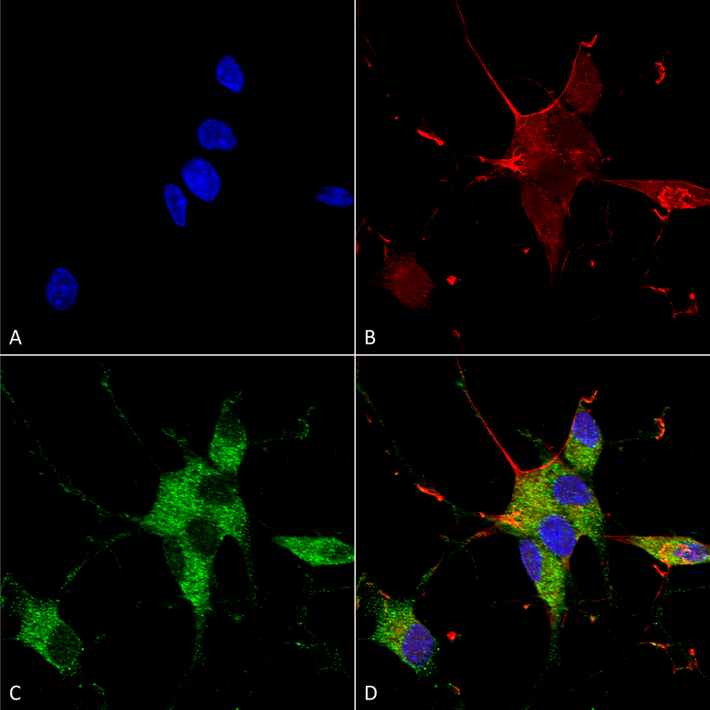 Immunocytochemistry/Immunofluorescence analysis using Mouse Anti-SHANK1/SHANK3 Monoclonal Antibody, Clone S367-51 (56578). Tissue: Neuroblastoma cells (SH-SY5Y). Species: Human. Fixation: 4% PFA for 15 min. Primary Antibody: Mouse Anti-SHANK1/SHANK3 Monoclonal Antibody (56578) at 1:100 for overnight at 4°C with slow rocking. Secondary Antibody: AlexaFluor 488 at 1:1000 for 1 hour at RT. Counterstain: Phalloidin-iFluor 647 (red) F-Actin stain; Hoechst (blue) nuclear stain at 1:800, 1.6mM for 20 min at RT. (A) Hoechst (blue) nuclear stain. (B) Phalloidin-iFluor 647 (red) F-Actin stain. (C) SHANK1/SHANK3 Antibody (D) Composite.
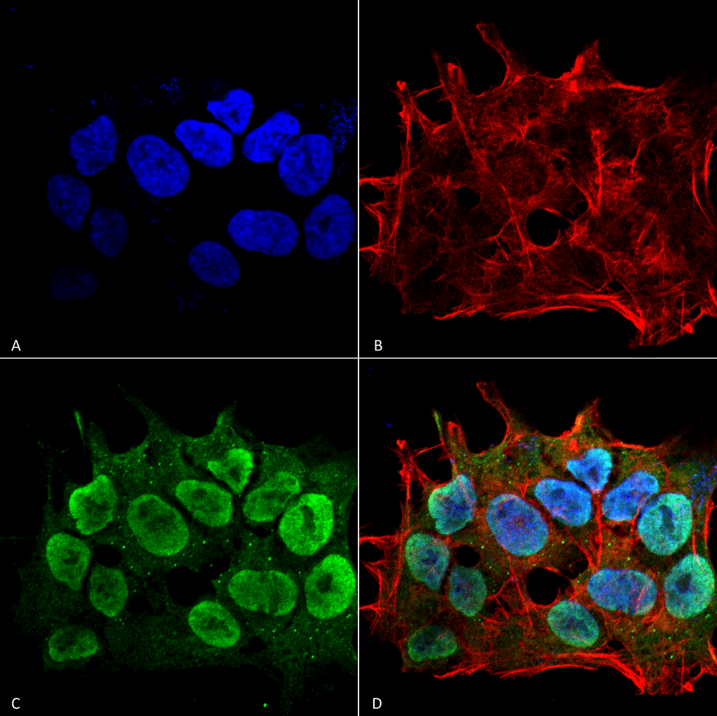 Immunocytochemistry/Immunofluorescence analysis using Mouse Anti-SHANK1/SHANK3 Monoclonal Antibody, Clone S367-51 (56578). Tissue: Neuroblastoma cell line (SK-N-BE). Species: Human. Fixation: 4% Formaldehyde for 15 min at RT. Primary Antibody: Mouse Anti-SHANK1/SHANK3 Monoclonal Antibody (56578) at 1:100 for 60 min at RT. Secondary Antibody: Goat Anti-Mouse ATTO 488 at 1:100 for 60 min at RT. Counterstain: Phalloidin Texas Red F-Actin stain; DAPI (blue) nuclear stain at 1:1000; 1:5000 for 60 min RT, 5 min RT. Localization: Cytoplasm, Nucleus. Magnification: 60X. (A) DAPI (blue) nuclear stain. (B) Phalloidin Texas Red F-Actin stain. (C) SHANK1/SHANK3 Antibody. (D) Composite.
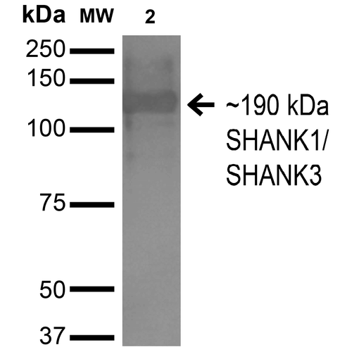 Western Blot analysis of Monkey COS cells transfected with HA-tagged Shank1 showing detection of ~190 kDa SHANK1/SHANK3 protein using Mouse Anti-SHANK1/SHANK3 Monoclonal Antibody, Clone S367-51 (56578). Lane 1: Molecular Weight Ladder. Lane 2: Monkey COS cells transfected with HA-tagged Shank1. Load: 15 µg. Block: 2% BSA and 2% Skim Milk in 1X TBST. Primary Antibody: Mouse Anti-SHANK1/SHANK3 Monoclonal Antibody (56578) at 1:200 for 16 hours at 4°C. Secondary Antibody: Goat Anti-Mouse IgG: HRP at 1:1000 for 1 hour RT. Color Development: ECL solution for 6 min in RT. Predicted/Observed Size: ~190 kDa.
 - -
- -
Antibody DetailsProduct DetailsReactive Species Human ⋅ Mouse ⋅ Rat Host Species Mouse Immunogen Fusion protein corresponding to aa 538-626 (SH3 domain) of rat SHANK3. This sequence is 100 Product Concentration 1.0 mg/ml Formulation PBS, pH 7.4, 0.1% sodium azide, 50% glycerol. State of Matter Liquid Product Preparation Purified by Protein G affinity chromatography Storage and Handling This product is stable for at least one (1) year at -20°C. Regulatory Status For in vitro investigational use only. Not intended for therapeutic or diagnostic procedures. Country of Origin USA Shipping Next Day 2-8°C Applications and Recommended Usage? Quality Tested by Leinco Immunofluorescence: use at 10ug/ml. These are recommended concentrations. Endusers should determine optimal concentrations for their application. Each investigator should determine their own optimal working dilution for specific applications. See directions on lot specific datasheets, as information may periodically change. DescriptionDescriptionSpecificity This antibody recognizes human, mouse, and rat SHANK1/SHANK3. Background SHANK proteins are scaffolding adaptors that have been shown to integrate neurotransmitter receptors into the cortical cytoskeleton at postsynaptic densities. SHANK1-3 of the SHANK/ProSAP family are molecular scaffolds in the postsynaptic density (PSD). SHANK recruits betaPIX and PAK to dendritic spines to regulate postsynaptic structure and interacts with ionotropic receptor and metabotropic glutamate receptor complexes. Transcript splice variation in the Shank family influences the spectrum of Shank-interacting proteins in the PSDs of adult and developing brain to ensure normal development. Function Major scaffold postsynaptic density protein which interacts with multiple proteins and complexes to orchestrate the dendritic spine and synapse formation, maturation and maintenance. Interconnects receptors of the postsynaptic membrane including NMDA-type and metabotropic glutamate receptors via complexes with GKAP/PSD-95 and HOMER, respectively, and the actin-based cytoskeleton. Plays a role in the structural and functional organization of the dendritic spine and synaptic junction through the interaction with Arp2/3 and WAVE1 complex as well as the promotion of the F-actin clusters. By way of this control of actin dynamics, participates in the regulation of developing neurons growth cone motility and the NMDA receptor-signaling. Also modulates GRIA1 exocytosis and GRM5/MGLUR5 expression and signaling to control the AMPA and metabotropic glutamate receptor-mediated synaptic transmission and plasticity. May be required at an early stage of synapse formation and be inhibited by IGF1 to promote synapse maturation. {PubMed:21606927, PubMed:21795692, PubMed:23100419, PubMed:23897824, PubMed:24089484}. NCBI Gene Bank ID UniProt.org Research Area Neuroscience References & CitationsTechnical Protocols  ICC/IF Certificate of Analysis |
Formats Available
 Products are for research use only. Not for use in diagnostic or therapeutic procedures.