

Anti-TrpV3 Cation Channel Antibody (11527)

Anti-TrpV3 Cation Channel Antibody (11527)
Product No.: 11527
- -
- -
Clone S15-39 Target TrpV3 Cation Channel Formats AvailableView All Product Type Monoclonal Isotype Mouse IgG1 Applications IHC , IP , WB , AM , ICC/IF |
Data
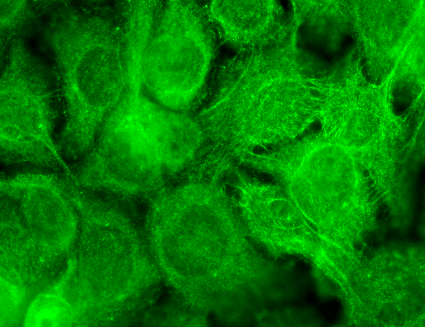 Immunocytochemistry/Immunofluorescence analysis using Mouse Anti-TrpV3 Monoclonal Antibody, Clone S15-39 (11527). Tissue: HaCaT cells. Species: Human. Fixation: Cold 100% methanol for 10 minutes at -20°C. Primary Antibody: Mouse Anti-TrpV3 Monoclonal Antibody (11527) at 1:100 for 1 hour at RT. Secondary Antibody: FITC Goat Anti-Mouse (green) at 1:50 for 1 hour at RT. Localization: Dotty staining in all cells. Some intermediate filament-like staining in some cells.
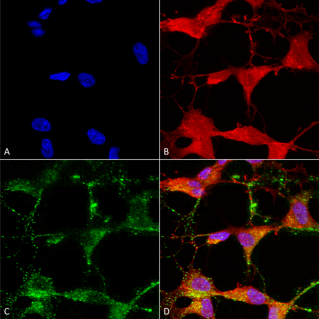 Immunocytochemistry/Immunofluorescence analysis using Mouse Anti-TrpV3 Monoclonal Antibody, Clone S15-39 (11527). Tissue: Neuroblastoma cells (SH-SY5Y). Species: Human. Fixation: 4% PFA for 15 min. Primary Antibody: Mouse Anti-TrpV3 Monoclonal Antibody (11527) at 1:50 for overnight at 4°C with slow rocking. Secondary Antibody: AlexaFluor 488 at 1:1000 for 1 hour at RT. Counterstain: Phalloidin-iFluor 647 (red) F-Actin stain; Hoechst (blue) nuclear stain at 1:800, 1.6mM for 20 min at RT. (A) Hoechst (blue) nuclear stain. (B) Phalloidin-iFluor 647 (red) F-Actin stain. (C) TrpV3 Antibody (D) Composite.
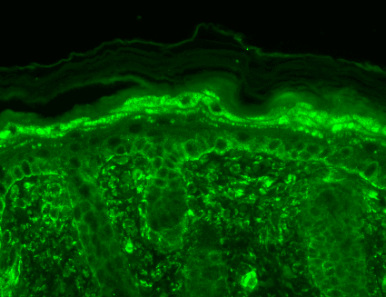 Immunohistochemistry analysis using Mouse Anti-TrpV3 Monoclonal Antibody, Clone S15-39 (11527). Tissue: backskin. Species: Mouse. Fixation: Bouin’s Fixative and paraffin-embedded. Primary Antibody: Mouse Anti-TrpV3 Monoclonal Antibody (11527) at 1:100 for 1 hour at RT. Secondary Antibody: FITC Goat Anti-Mouse (green) at 1:50 for 1 hour at RT. Localization: Filaggrin-like staining in upper layers. Dull lower layer cell staining. Some stain seen in hypodermis.
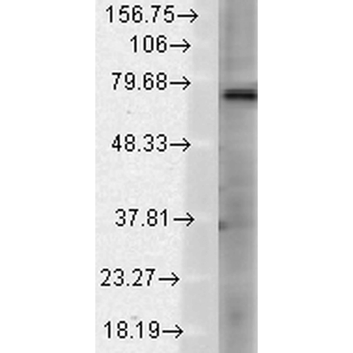 Western Blot analysis of Rat brain membrane lysate showing detection of TrpV3 protein using Mouse Anti-TrpV3 Monoclonal Antibody, Clone S15-39 (11527). Load: 15 µg. Block: 1.5% BSA for 30 minutes at RT. Primary Antibody: Mouse Anti-TrpV3 Monoclonal Antibody (11527) at 1:1000 for 2 hours at RT. Secondary Antibody: Sheep Anti-Mouse IgG: HRP for 1 hour at RT.
 - -
- -
Antibody DetailsProduct DetailsReactivity Species Human ⋅ Mouse ⋅ Rat Host Species Mouse Immunogen Synthetic peptide corresponding to aa 458-474 (cytoplasmic C-terminus) of rat TrpV3 (accession no. NP_001020928). This sequence is 77 Product Concentration Lot Specific Formulation PBS, pH 7.4; 50% glycerol, 0.09% sodium azide. State of Matter Liquid Product Preparation Purified by Protein G affinity chromatography Storage and Handling This antibody is stable for at least one (1) year at -20°C. Avoid repeated freeze-thaw cycles. Regulatory Status For in vitro investigational use only. Not for use in therapeutic or diagnostic procedures. Country of Origin USA Shipping Next Day 2-8°C Applications and Recommended Usage? Quality Tested by Leinco Immunoblotting: use at 1ug/mL. A band of ~70kDa is detected.Immunohistochemistry and
Immunocytochemistry: use at 0.1-1ug/mL Immunofluorescence: use at 1-10ug/mL These are recommended concentrations. User should determine optimal concentrations for their application. Positive control: Lysate of COS-1 cells transiently-transfected with TrpV3. Each investigator should determine their own optimal working dilution for specific applications. See directions on lot specific datasheets, as information may periodically change. DescriptionSpecificity This antibody recognizes human, mouse, and rat TrpV3. Background Ion channels are integral membrane proteins that help establish and control the small voltage gradient across the plasma membrane of living cells by allowing the flow of ions down their electrochemical gradient. TrpV3 is in a family of non- selective cation channels that function in a variety of processes including temperature sensation and vaso-regulation. The thermosensitive members of this family are expressed in sensory neurons that terminate in the skin and is activated at temperatures between 22-40oC. TrpV3 might associate with TrpV1 to form heteromeric channels. Antigen DetailsUniProt.org Research Area Ion Channels References & CitationsTechnical Protocols   AM ICC/IF |
