

Anti-VDAC1 Antibody

Anti-VDAC1 Antibody
Product No.: 56564
- -
- -
Clone S152B-23 Target VDAC1 Formats AvailableView All Product Type Monoclonal Alternate Names VDAC-1, hVDAC1, Outer mitochondrial membrane protein porin 1, Plasmalemmal porin, Porin 31HL, Porin 31HM Isotype Mouse IgG2a Applications ICC , IF , IHC , WB |
Data
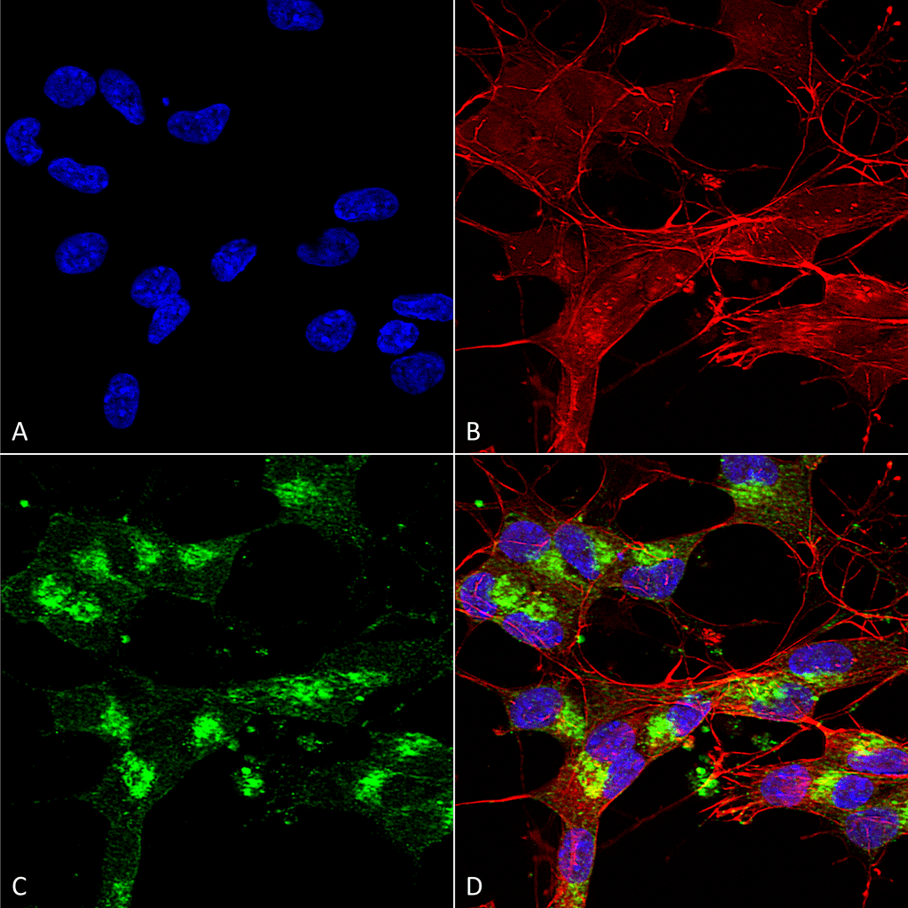 Immunocytochemistry/Immunofluorescence analysis using Mouse Anti-VDAC1 Monoclonal Antibody, Clone S152B-23 (56564). Tissue: Neuroblastoma cells (SH-SY5Y). Species: Human. Fixation: 4% PFA for 15 min. Primary Antibody: Mouse Anti-VDAC1 Monoclonal Antibody (56564) at 1:100 for overnight at 4°C with slow rocking. Secondary Antibody: AlexaFluor 488 at 1:1000 for 1 hour at RT. Counterstain: Phalloidin-iFluor 647 (red) F-Actin stain; Hoechst (blue) nuclear stain at 1:800, 1.6mM for 20 min at RT. (A) Hoechst (blue) nuclear stain. (B) Phalloidin-iFluor 647 (red) F-Actin stain. (C) VDAC1 Antibody (D) Composite.
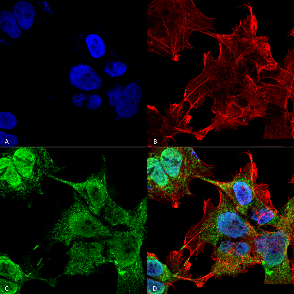 Immunocytochemistry/Immunofluorescence analysis using Mouse Anti-VDAC1 Monoclonal Antibody, Clone S152B-23 (56564). Tissue: Neuroblastoma cell line (SK-N-BE). Species: Human. Fixation: 4% Formaldehyde for 15 min at RT. Primary Antibody: Mouse Anti-VDAC1 Monoclonal Antibody (56564) at 1:100 for 60 min at RT. Secondary Antibody: Goat Anti-Mouse ATTO 488 at 1:100 for 60 min at RT. Counterstain: Phalloidin Texas Red F-Actin stain; DAPI (blue) nuclear stain at 1:1000; 1:5000 for 60 min RT, 5 min RT. Localization: Mitochondrion, Mitochondrion Outer Membrane, Nucleus. Magnification: 60X. (A) DAPI (blue) nuclear stain. (B) Phalloidin Texas Red F-Actin stain. (C) VDAC1 Antibody. (D) Composite.
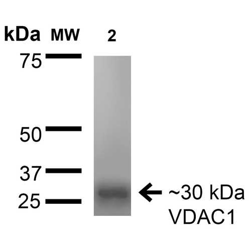 Western Blot analysis of Rat Brain Membrane showing detection of ~30 kDa VDAC1 protein using Mouse Anti-VDAC1 Monoclonal Antibody, Clone S152B-23 (56564). Lane 1: Molecular Weight Ladder. Lane 2: Rat Brain Membrane. Load: 15 µg. Block: 2% BSA and 2% Skim Milk in 1X TBST. Primary Antibody: Mouse Anti-VDAC1 Monoclonal Antibody (56564) at 1:200 for 16 hours at 4°C. Secondary Antibody: Goat Anti-Mouse IgG: HRP at 1:1000 for 1 hour RT. Color Development: ECL solution for 6 min in RT. Predicted/Observed Size: ~30 kDa.
 - -
- -
Antibody DetailsProduct DetailsReactive Species Human ⋅ Mouse ⋅ Rat Host Species Mouse Immunogen Fusion protein corresponding to aa 1-283 (full-length) of human VDAC1. This sequence is 98 Product Concentration 1.0 mg/ml Formulation PBS, pH 7.4, 0.1% sodium azide, 50% glycerol. State of Matter Liquid Product Preparation Purified by Protein G affinity chromatography Storage and Handling This product is stable for at least one (1) year at -20°C. Regulatory Status For in vitro investigational use only. Not intended for therapeutic or diagnostic procedures. Country of Origin USA Shipping Next Day 2-8°C Applications and Recommended Usage? Quality Tested by Leinco Immunoblotting: use at 1-5ug/mL. A band of ~30kDa is detected.
Immunofluorescence: use at 10ug/mL These are recommended concentrations. Endusers should determine optimal concentrations for their application. Each investigator should determine their own optimal working dilution for specific applications. See directions on lot specific datasheets, as information may periodically change. DescriptionDescriptionSpecificity This antibody recognizes human, mouse and rat VDAC1. It does not cross-react
with VDAC2 or VDAC3 (based on evaluation in knock-out mice). Background VDAC1 (also known as Voltage-dependent anion-selective channel protein 1 and outer mitochondrial membrane protein porin 1) is the outer mitochondrial membrane receptor for hexokinase and BCL2L1. VDAC1 forms a channel through the mitochondrial membrane and is involved in small molecule diffusion, cell volume regulation, and apoptosis. VDAC1 may participate in the formation of the permeability transition pore complex (PTPC) which is responsible for the release of mitochondrial products that trigger apoptosis. Function Forms a channel through the mitochondrial outer membrane and also the plasma membrane. The channel at the outer mitochondrial membrane allows diffusion of small hydrophilic molecules; in the plasma membrane it is involved in cell volume regulation and apoptosis. It adopts an open conformation at low or zero membrane potential and a closed conformation at potentials above 30-40 mV. The open state has a weak anion selectivity whereas the closed state is cation-selective (PubMed:11845315, PubMed:18755977, PubMed:20230784, PubMed:8420959). Binds various signaling molecules, including the sphingolipid ceramide, the phospholipid phosphatidylcholine, and the sterol cholesterol (PubMed:31015432). In depolarized mitochondria, acts downstream of PRKN and PINK1 to promote mitophagy or prevent apoptosis; polyubiquitination by PRKN promotes mitophagy, while monoubiquitination by PRKN decreases mitochondrial calcium influx which ultimately inhibits apoptosis (PubMed:32047033). May participate in the formation of the permeability transition pore complex (PTPC) responsible for the release of mitochondrial products that triggers apoptosis (PubMed:15033708, PubMed:25296756). May mediate ATP export from cells (PubMed:30061676). {PubMed:11845315, PubMed:15033708, PubMed:18755977, PubMed:20230784, PubMed:25296756, PubMed:30061676, PubMed:31015432, PubMed:32047033, PubMed:8420959}. NCBI Gene Bank ID UniProt.org Research Area Neuroscience References & CitationsTechnical ProtocolsICC IF   Certificate of Analysis |
