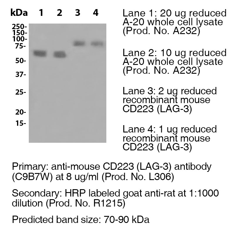
mAbMods™ Anti-Mouse CD223 (LAG-3) Antibody (Clone C9B7W)
mAbMods™ Anti-Mouse CD223 (LAG-3) Antibody (Clone C9B7W)
Product No.: L313
- -
- -
Product No.L313 Clone C9B7W Target CD223 Formats AvailableView All Product Type Recombinant Monoclonal Antibody for in vivo Use Alternate Names CD223, LAG3 Isotype Mouse IgG1 Applications B , FA , FC , in vivo , IP , WB |
Data
- -
- -
Antibody DetailsProduct DetailsReactive Species Mouse Host Species Mouse-Rat Chimeric FC Effector Activity Active Recommended Isotype Controls Recommended Dilution Buffer Immunogen Murine CD223-Ig fusion protein Product Concentration ≥ 5.0 mg/ml Endotoxin Level <0.5 EU/mg as determined by the LAL method Purity ≥95% by SDS Page ⋅ ≥95% monomer by analytical SEC Formulation This recombinant monoclonal antibody is aseptically packaged and formulated in 0.01 M phosphate buffered saline (150 mM NaCl) PBS pH 7.2 - 7.4 with no carrier protein, potassium, calcium or preservatives added. Due to inherent biochemical properties of antibodies, certain products may be prone to precipitation over time. Precipitation may be removed by aseptic centrifugation and/or filtration. State of Matter Liquid Product Preparation Functional grade preclinical antibodies are manufactured in an animal free facility using in vitro cell culture techniques and are purified by a multi-step process including the use of protein A or G to assure extremely low levels of endotoxins, leachable protein A or aggregates. Pathogen Testing To protect mouse colonies from infection by pathogens and to assure that experimental preclinical data is not affected by such pathogens, all of Leinco’s recombinant antibodies are tested and guaranteed to be negative for all pathogens in the IDEXX IMPACT I Mouse Profile. Storage and Handling Functional grade preclinical antibodies may be stored sterile as received at 2-8°C for up to one month. For longer term storage, aseptically aliquot in working volumes without diluting and store at ≤ -70°C. Avoid Repeated Freeze Thaw Cycles. Country of Origin USA Shipping Next Day 2-8°C RRIDAB_2737552 Additional Applications Reported In Literature ? B LAG-3 antibody (clone C9B7W) blocks the function of murine LAG-3 in vivo. Each investigator should determine their own optimal working dilution for specific applications. See directions on lot specific datasheets, as information may periodically change. DescriptionDescriptionSpecificity LAG-3 antibody (clone C9B7W) recognizes and specifically binds to an epitope in the D2 domain of CD223. Background This anti-Lag-3 antibody is a recombinant chimeric version of the original clone C9B7W antibody. The variable domain sequences are identical to the original clone C9B7W; however, the constant region sequences have been switched from rat IgG1 to mouse IgG1.
Lymphocyte activation gene (LAG)-3 is a CD4-related cell surface molecule that binds to MHC class II molecules and acts as a negative regulator of T cell function by limiting the expansion of activated T cells and T cell homeostasis 1,2,3. LAG-3 inhibits CD4-dependent T cell function via its cytoplasmic domain and a conserved “KIEELE” motif is essential for interaction with downstream signaling molecules 2. Murine LAG-3 RNA is strongly expressed in the thymus, spleen, and areas of the brain outside the cerebellum as well as in day 7 postnatal brain at the base of the cerebellum, developing white matter and choroid plexus. However, LAG-3 protein is detected on the cell surface of a very small percentage of T and natural killer cells in naïve mice, meaning RNA abundance is not indicative of protein levels for this gene. The original clone C9B7W was generated by immunizing Lewis rats with an mLAG-3:Ig fusion protein consisting of the entire extracellular domain of LAG-3 attached to the murine IgG1 heavy chain 1. C9B7W recognizes an epitope in the D2 domain of LAG-3 and does not block binding of MHC class II:γ2aFc-Alexa633 to murine LAG-3+ T cell hybridomas. However, C9B7W does inhibit antigen-induced IL-2 production. Antigen Distribution LAG-3 is uniformly expressed by antigen-activated T cells. LAG-3 protein is expressed on the cell surface of a very small percentage of T and natural killer cells in naïve mice. These cells include γ δ T cells, dividing memory T cells, CD8+, and CD4+ T cells. NCBI Gene Bank ID UniProt.org Research Area Cell Biology . Immunology . Inhibitory Molecules References & Citations1. Workman CJ, Rice DS, Dugger KJ, et al. Eur J Immunol. 32(8):2255-2263. 2002. 2. Workman CJ, Dugger KJ, Vignali DA. J Immunol. 169(10):5392-5395. 2002. 3. Workman CJ, Vignali DA. J Immunol. 174(2):688-695. 2005. 4. Li N, Workman CJ, Martin SM, et al. J Immunol. 173(11):6806-6812. 2004. Technical ProtocolsCertificate of Analysis |
Related Products
- -
- -
Prod No. | Description |
|---|---|
S211 | |
R1364 | |
I-1195 | |
C247 | |
F1175 | |
R1214 | |
S571 |
Formats Available
- -
- -
Prod No. | Description |
|---|---|
C2252 | |
C2253 | |
C2254 | |
C2255 | |
C2256 | |
C2249 | |
C2251 | |
C2250 | |
L306 | |
C2155 | |
L502 | |
C2852 | |
L313 | |
L310 |


